Summary: Researchers reveal in mice, certain cells within the hippocampus fire when the animal is anxious and this triggers anxiety related behaviors to occur.
Source: Columbia University Medical Center.
Do your palms sweat when you walk down a poorly lit street at night? That feeling may be traced to the firing of newly identified “anxiety” cells deep inside your brain, according to new research from neuroscientists at Columbia University Irving Medical Center (CUIMC) and the University of California, San Francisco (UCSF).
The researchers found the cells in the brains of mice, inside a structure called the hippocampus. But the cells probably also exist in humans, says Rene Hen, PhD, a professor of psychiatry at CUIMC and one of the study’s senior investigators.
“We call these anxiety cells because they only fire when the animals are in places that are innately frightening to them,” Hen says. “For a mouse, that’s an open area where they’re more exposed to predators, or an elevated platform.”
The firing of the anxiety cells sends messages to other parts of the brain that turn on anxious behaviors–in mice, those include avoiding the dangerous area or fleeing to a safe zone.
Though many other cells in the brain have been identified as playing a role in anxiety, the cells found in this study are the first known to represent the state of anxiety, regardless of the type of environment that provokes the emotion.
“This is exciting because it represents a direct, rapid pathway in the brain that lets animals respond to anxiety-provoking places without needing to go through higher-order brain regions,” said Mazen Kheirbek, PhD, an assistant professor of psychiatry at UCSF and study’s other senior investigator.
“Now that we’ve found these cells in the hippocampus, it opens up new areas for exploring treatment ideas that we didn’t know existed before,” says the study’s lead author Jessica Jimenez, PhD, an MD/PhD student at Columbia University’s Vagelos College of Physicians & Surgeons.
The findings appear in the January 31 online issue of Neuron. “This study shows how translational research using basic science techniques in animal models can elucidate the underlying basis of human emotions and reasons for mental disorders, thereby pointing the way for treatment development,” says Jeffrey Lieberman, MD, Lawrence C. Kolb Professor and Chair of Psychiatry at CUIMC.
Anxiety is healthy. To a degree.
Anxiety is normal and critical to an animal’s safety. Anxiety is an emotional response to a distant threat–being in an environment that exposes an animal to predators, for example. The safe bet is to sidestep those environments, so anxiety kicks in avoidance behaviors.
When people overestimate threats–when talking to a crowd invokes the same response as potentially running into a snake–anxiety becomes a problem.
To understand how things go awry in anxiety disorders, researchers in the Hen lab have been looking at mice to decipher how the brain processes healthy anxiety.
“We wanted to understand where the emotional information that goes into the feeling of anxiety is encoded within the brain,” said Mazen Kheirbek, PhD, who was an assistant professor at CUIMC before moving to UCSF.
The hippocampus plays a well-known role in the brain’s ability to form new memories and to help animals–from mice to humans–navigate through complex environments. But recent research has also implicated the hippocampus in regulating mood, and studies have shown altering brain activity in the ventral part of the hippocampus can reduce anxiety. It’s also known that the hippocampus sends signals to other areas of the brain–the amygdala and the hypothalamus–that have also been shown to control anxiety-related behavior.
Anxiety cells identified with miniature microscope
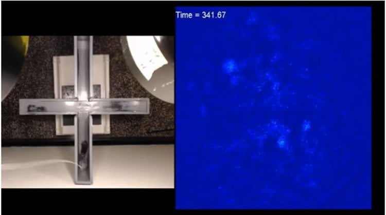
Using a miniature microscope inserted into the brains of the mice, Hen’s team recorded the activity of hundreds of cells in the hippocampus as the mice freely moved around their surroundings.
Whenever the animals were in exposed, anxiety-provoking environments, the researchers noticed that specific cells in the ventral part of the hippocampus were active. And the more anxious the mice seemed, the greater the activity in the cells.
The researchers traced the output of those cells to the hypothalamus, which is known to control behaviors associated with anxiety (in people, those include increased heart rate, avoidance, and secretion of stress hormones).
By turning the anxiety cells off and on using a technique called optogenetics that allows scientists to control the activity of neurons using beams of light, the researchers found that the anxiety cells control anxiety behaviors. When the cells were silenced, the mice stopped producing fear-related behaviors, wandering onto elevated platforms and away from protective walls. When the anxiety cells were stimulated, the mice exhibited more fear behaviors even when they were in “safe” surroundings.
Therapeutic directions
The discovery of the anxiety cells raises the possibility of finding treatments that target them and reduce anxiety. “We’re looking to see if these cells are different molecularly from other neurons,” Hen says. “If there’s a specific receptor on the cells that distinguishes them from their neighbors, it may be possible to produce a new drug to reduce anxiety.”
Funding: The study was supported by grants from the National Institutes of Health (KO1AG054765, R90DA023426, 2RO1MH064537, R01EB22913, R37MH068542, R01AG043688, R01MH083862, R01MH108623, R01MH111754), Intelligence Advanced Research Projects Activity, Defense Advanced Research Projects Agency, NY STEM awards, and the Weill Scholar Award program.
Additional authors are Jessica C. Jimenez (Columbia University Irving Medical Center, New York, NY), Katy Su (CUIMC), Alexander R. Goldberg (CUIMC), Victor M. Luna (CUIMC), Jeremy S. Biane (University of California, San Francisco, CA), Gokhan Ordek (CUIMC), Pengcheng Zhou (Carnegie Mellon University, Pittsburgh, PA), Samantha K. Ong (CUIMC), Matthew A. Wright (Stanford University, Palo Alto, CA), Larry Zweifel (University of Washington, Seattle, WA), and Liam Peninski (Columbia University).
The authors report no financial or other conflicts of interest.
Source: Helen Garey – Columbia University Medical Center
Publisher: Organized by NeuroscienceNews.com.
Image Source: NeuroscienceNews.com image is credited to the lab of Rene Hen, PhD, Columbia University Irving Medical Center.
Original Research: Study will appear in Neuron.
[cbtabs][cbtab title=”MLA”]Columbia University Medical Center “‘Anxiety Cells’ Identified in the Hippocampus.” NeuroscienceNews. NeuroscienceNews, 31 January 2018.
<https://neurosciencenews.com/hippocampus-anxiety-cells-8408/>.[/cbtab][cbtab title=”APA”]Columbia University Medical Center (2018, January 31). ‘Anxiety Cells’ Identified in the Hippocampus. NeuroscienceNews. Retrieved January 31, 2018 from https://neurosciencenews.com/hippocampus-anxiety-cells-8408/[/cbtab][cbtab title=”Chicago”]Columbia University Medical Center “‘Anxiety Cells’ Identified in the Hippocampus.” https://neurosciencenews.com/hippocampus-anxiety-cells-8408/ (accessed January 31, 2018).[/cbtab][/cbtabs]