

Summary: Findings shed light on the mechanisms underlying brain damage in microgravity.
Source: Science China Press
Although they have been investigated for the last decade, the mechanisms that underlie microgravity-induced neurological changes have not yet fully been elucidated. Upward fluid transfer following exposure to microgravity leads to certain adaptive changes in the brain parenchyma, cerebrospinal fluid (CSF), and blood vessels.
The brain extracellular space (ECS) contains brain interstitial fluid (ISF) and occupies 15%–20% of the living brain by volume. Since the brain ECS serves as an important intermediate station for material transfer among capillaries, neural cells, and CSF, the alterations to this space that occur following exposure to microgravity remain unclear.
In this study, the brain ECS and drainage of the ISF under simulated microgravity were investigated using a self-developed measuring instrument with Decs-mapping technique. Changes of ECS and substance transportation within were demonstrated at very early stage.
Drainage of ISF was accelerated at day 3 and slowed at day 7. There was no abnormal neuronal morphology detected by HE staining and TUNEL in the HU-3 group; the percentages of hyperchromatic nuclei and apoptotic neurons showed no difference from the CON-3 group. However, in the HU-7 group, the abnormal cell structure associated with an increase in the apoptosis of cells did not recover in the hippocampal CA1 region.
After the reloading process, the faster drainage at day 3 returned to the normal level, while the slow drainage at day 7 was irreversible. The brain ISF flows in the ECS and its drainage plays a significant role in maintaining local homeostasis for neuronal activities. Actually, ISF drainage is affected by both the local properties of the brain ECS and the drainage pathway.
These findings provided a new way to understand the mechanism underlying potential brain damage under microgravity, as well as a perspective from which we may be able to protect astronauts.
Both ISF drainage and ECS diffusion could be regulated by neurotransmitters released following neuronal activities.
Meanwhile, the exploration of mutual interactions between the neural networks, the brain ECS, and the vascular system is worthy of further investigation. Elucidating the ECS and ISF changes over a longer duration of simulated microgravity will contribute to the protection of astronauts in upcoming long-term manned space flights or moon landing missions.
About this neuroscience research news
Author: Bei Yan
Source: Science China Press
Contact: Bei Yan – Science China Press
Image: The image is credited to Science China Press
Original Research: Closed access.
“Early changes to the extracellular space in the hippocampus under simulated microgravity conditions” by Gao, Y., Han, H., Du, J.,et al. Science China Press Sciences
Abstract
Early changes to the extracellular space in the hippocampus under simulated microgravity conditions
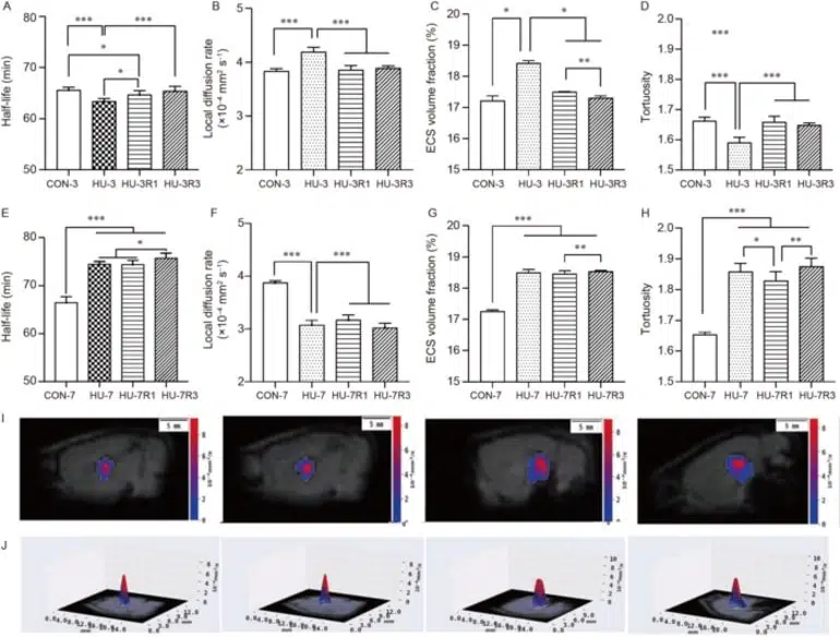
The smooth transportation of substances through the brain extracellular space (ECS) is crucial to maintaining brain function; however, the way this occurs under simulated microgravity remains unclear. In this study, tracer-based magnetic resonance imaging (MRI) and DECS-mapping techniques were used to image the drainage of brain interstitial fluid (ISF) from the ECS of the hippocampus in a tail-suspended hindlimb-unloading rat model at day 3 (HU-3) and 7 (HU-7).
The results indicated that drainage of the ISF was accelerated in the HU-3 group but slowed markedly in the HU-7 group. The tortuosity of the ECS decreased in the HU-3 group but increased in the HU-7 group, while the volume fraction of the ECS increased in both groups.
The diffusion rate within the ECS increased in the HU-3 group and decreased in the HU-7 group. The alterations to ISF drainage and diffusion in the ECS were recoverable in the HU-3 group, but neither parameter was restored in the HU-7 group.
Our findings suggest that early changes to the hippocampal ECS and ISF drainage under simulated microgravity can be detected by tracer-based MRI, providing a new perspective for studying microgravity-induced nano-scale structure abnormities and developing neuroprotective approaches involving the brain ECS.


