

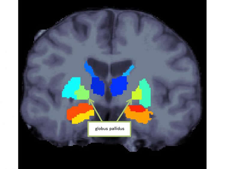
A research group led by Ryota Hashimoto, an associate professor at Osaka University, Naohiro Okada, a graduate student at the University of Tokyo, and Kiyoto Kasai, a professor at the University of Tokyo, replicated prior findings that the volume of globus pallidus (one of the basal ganglia in the brain) in schizophrenia was larger than that in healthy subjects. Also, the group found that patients with schizophrenia demonstrated a specific leftward volumetric asymmetry for the globus pallidus.
They compared and analyzed magnetic resonance imaging (MRI) brain images of 1,680 healthy individuals and 884 patients with schizophrenia from 11 research institutes participating in Cognitive Genetics Collaborative Research Organization (COCORO), and examined the differences between schizophrenia and healthy controls in the subcortical regional volumes and their asymmetries.
Compared to controls, patients with schizophrenia demonstrated smaller bilateral hippocampus, amygdala, thalamus and accumbens volumes as well as intracranial volume, but larger bilateral caudate, putamen, pallidum and lateral ventricle volumes. Also, patients with schizophrenia demonstrated a specific leftward asymmetry for globus pallidus volume.
These results suggest the possibility of aberrant laterality in neural pathways and connectivity patterns related to the globus pallidus in schizophrenia. Further, through the elucidation of the underlying pathological mechanisms, it will be a step toward the development of therapeutic strategies for schizophrenia.
Funding: Funding was provided by Japan Agency for Medical Research and Development, Ministry of Education, Culture, Sports, Science and Technology, Japan.
Source: Ryota Hashimoto – Osaka University
Image Source: The image is credited to Osaka University.
Original Research: Full open access research for “Abnormal asymmetries in subcortical brain volume in schizophrenia” by N Okada, M Fukunaga, F Yamashita, D Koshiyama, H Yamamori, K Ohi, Y Yasuda, M Fujimoto, N Yahata, K Nemoto, D P Hibar, T G M van Erp, H Fujino, M Isobe, S Isomura, T Natsubori, H Narita, N Hashimoto, J Miyata, S Koike, T Takahashi, H Yamasue, K Matsuo, T Onitsuka, T Iidaka, Y Kawasaki, R Yoshimura, M Suzuki, J A Turner, M Takeda, P M Thompson, N Ozaki, K Kasai and R Hashimoto in Molecular Psychiatry. Published online January 191 2016 doi:10.1038/mp.2015.209
Abstract
Abnormal asymmetries in subcortical brain volume in schizophrenia
Subcortical structures, which include the basal ganglia and parts of the limbic system, have key roles in learning, motor control and emotion, but also contribute to higher-order executive functions. Prior studies have reported volumetric alterations in subcortical regions in schizophrenia. Reported results have sometimes been heterogeneous, and few large-scale investigations have been conducted. Moreover, few large-scale studies have assessed asymmetries of subcortical volumes in schizophrenia. Here, as a work completely independent of a study performed by the ENIGMA consortium, we conducted a large-scale multisite study of subcortical volumetric differences between patients with schizophrenia and controls. We also explored the laterality of subcortical regions to identify characteristic similarities and differences between them. T1-weighted images from 1680 healthy individuals and 884 patients with schizophrenia, obtained with 15 imaging protocols at 11 sites, were processed with FreeSurfer. Group differences were calculated for each protocol and meta-analyzed. Compared with controls, patients with schizophrenia demonstrated smaller bilateral hippocampus, amygdala, thalamus and accumbens volumes as well as intracranial volume, but larger bilateral caudate, putamen, pallidum and lateral ventricle volumes. We replicated the rank order of effect sizes for subcortical volumetric changes in schizophrenia reported by the ENIGMA consortium. Further, we revealed leftward asymmetry for thalamus, lateral ventricle, caudate and putamen volumes, and rightward asymmetry for amygdala and hippocampal volumes in both controls and patients with schizophrenia. Also, we demonstrated a schizophrenia-specific leftward asymmetry for pallidum volume. These findings suggest the possibility of aberrant laterality in neural pathways and connectivity patterns related to the pallidum in schizophrenia.
“Abnormal asymmetries in subcortical brain volume in schizophrenia” by N Okada, M Fukunaga, F Yamashita, D Koshiyama, H Yamamori, K Ohi, Y Yasuda, M Fujimoto, N Yahata, K Nemoto, D P Hibar, T G M van Erp, H Fujino, M Isobe, S Isomura, T Natsubori, H Narita, N Hashimoto, J Miyata, S Koike, T Takahashi, H Yamasue, K Matsuo, T Onitsuka, T Iidaka, Y Kawasaki, R Yoshimura, M Suzuki, J A Turner, M Takeda, P M Thompson, N Ozaki, K Kasai and R Hashimoto in Molecular Psychiatry. Published online January 191 2016 doi:10.1038/mp.2015.209


