Summary: According to a new study, people with schizophrenia show reduced levels of GABA and glutamate.
Source: Michigan State University.
The brains of healthy relatives of people with schizophrenia may hold a clue to better understand – and ultimately treat – the devastating illness, finds new research led by a Michigan State University scientist.
The study is the first to look at the neurotransmitters glutamate and gamma-aminobutyric acidergic, or GABA, in both schizophrenia patients and healthy relatives of schizophrenia patients using a noninvasive imaging test called magnetic resonance spectroscopy.
Glutamate, which promotes the firing of brain cells, and GABA, which inhibits neural firing, work hand in hand to regulate brain function. In the past 20 years, many researchers have come to believe that glutamate and GABA play a role in schizophrenia, yet the precise relationship remains unclear and no medication for schizophrenia has hit the market specifically targeting these neurotransmitters.
According to the study, both schizophrenia patients and healthy relatives showed reduced levels of glutamate. But while the patients also showed reduced levels of GABA, the relatives had normal amounts of the inhibitory neurotransmitter.
This begs two key questions. First, if glutamate is altered, why do these relatives not show symptoms of the illness? And, second, how did healthy relatives maintain normal levels of GABA even though they, like the patients, were genetically predisposed to schizophrenia and had altered glutamate levels?
“This finding is what’s most exciting about our study,” said lead investigator Katharine Thakkar, MSU assistant professor of clinical psychology. “It hints at what kinds of things have to go wrong for someone to express this vulnerability toward schizophrenia. The study gives us more specific clues into what kinds of systems we want to tackle when we’re developing new treatments for this very devastating illness.”
The research, reported online in the journal Biological Psychiatry, involved 21 patients with chronic schizophrenia, 23 healthy relatives (the relatives were siblings of other patients with schizophrenia, not the patients in the study) and a control group of 24 healthy nonrelatives. It was performed in collaboration with researchers at the University Medical Center Utrecht in the Netherlands, where Thakkar served as a postdoctoral fellow.
Schizophrenia, which affects about 3.5 million Americans, is marked by delusions, hallucinations and other symptoms, although it is not, contrary to popular belief, split or multiple personality.
Many schizophrenia drugs regulate dopamine, another neurotransmitter in the brain, though the medication does not work for everyone. In fact, medication for schizophrenia has changed very little in the past 50 years and remains somewhat limited in its effectiveness. Many researchers believe there are multiple risk factors for the illness, including dopamine and glutamate/GABA imbalance.
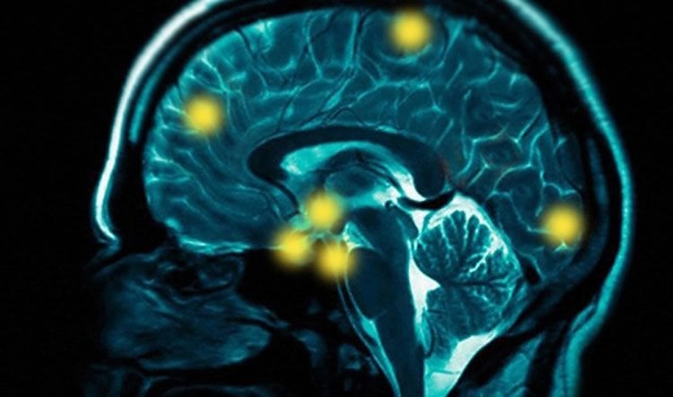
The brain scan used in the study – which is conducted on a conventional MRI machine – could eventually help clinicians target more specific treatments.
“There are likely different causes of the different symptoms and possibly different mechanisms of the illness across individuals,” Thakkar said. “In the future, as this imaging technique becomes more refined, it could conceivably be used to guide individual treatment recommendations. That is, this technique might indicate that one individual would benefit more from treatment A and another individual would benefit more from treatment B, when these different treatments have different mechanisms of action.”
Source: Andy Henion – Michigan State University
Image Source: This NeuroscienceNews.com image is adapted from the Michigan State University press release.
Original Research: Abstract for “7T proton magnetic resonance spectroscopy of GABA, glutamate, and glutamine reveals altered concentrations in schizophrenia patients and healthy siblings” by Katharine N. Thakkar, Lara Rösler, Jannie P. Wijnen, Vincent O. Boer, Dennis W.J. Klomp, Wiepke Cahn, René S. Kahn, and Sebastiaan F.W. Negger in Biological Psychiatry. Published online April 19 2016 doi:10.1016/j.biopsych.2016.04.007
[cbtabs][cbtab title=”MLA”]Michigan State University. “New Clues to Deciphering Schizophrenia.” NeuroscienceNews. NeuroscienceNews, 7 June 2016.
<https://neurosciencenews.com/gaba-schizophrenia-psychology-4395/>.[/cbtab][cbtab title=”APA”]Michigan State University. (2016, June 7). New Clues to Deciphering Schizophrenia. NeuroscienceNews. Retrieved June 7, 2016 from https://neurosciencenews.com/gaba-schizophrenia-psychology-4395/[/cbtab][cbtab title=”Chicago”]Michigan State University. “New Clues to Deciphering Schizophrenia.” https://neurosciencenews.com/gaba-schizophrenia-psychology-4395/ (accessed June 7, 2016).[/cbtab][/cbtabs]
Abstract
7T proton magnetic resonance spectroscopy of GABA, glutamate, and glutamine reveals altered concentrations in schizophrenia patients and healthy siblings
Background
The NMDA-receptor hypofunction model of schizophrenia predicts dysfunction in both glutamatergic and GABAergic transmission. We addressed this hypothesis by measuring γ-aminobutyric acid (GABA), glutamate (Glu), glutamine (Gln), and Glx (Gln+Glu) concentrations in vivo in patients with schizophrenia using proton magnetic resonance spectroscopy (1H-MRS) at 7 Tesla (7T), which allows separation of metabolites that would otherwise overlap at lower field strengths. In addition, we investigated whether altered levels of GABA, Glu, Gln, and Glx reflect genetic vulnerability towards schizophrenia by including healthy first-degree relatives.
Methods
21 chronic, medicated schizophrenia patients, 23 healthy first-degree relatives of schizophrenia patients, and 24 healthy non-relatives underwent 1H-MRS at 7T. Glu, Gln, and GABA were measured cortically and subcortically in bilateral basal ganglia and occipital cortex.
Results
Individuals with schizophrenia had reduced cortical GABA, compared with healthy relatives and the combined sample of healthy relatives and healthy non-relatives, suggesting that altered GABAergic systems in schizophrenia are associated with either disease state or medication effects. On the other hand, reduced cortical Glu relative to healthy controls was observed in schizophrenia patients and the combined sample of healthy relatives and schizophrenia patients, suggesting that altered glutamatergic metabolite levels are associated with illness liability. No group differences were found in the basal ganglia.
Conclusions
Combined, these findings are consistent with alterations in GABAergic and glutamatergic systems in schizophrenia and provide novel insights into these systems in healthy relatives.
“7T proton magnetic resonance spectroscopy of GABA, glutamate, and glutamine reveals altered concentrations in schizophrenia patients and healthy siblings” by Katharine N. Thakkar, Lara Rösler, Jannie P. Wijnen, Vincent O. Boer, Dennis W.J. Klomp, Wiepke Cahn, René S. Kahn, and Sebastiaan F.W. Negger in Biological Psychiatry. Published online April 19 2016 doi:10.1016/j.biopsych.2016.04.007