Summary: Commonly associated with helping improve brain function, the omega 3 fatty acid DHA may have another, previously unknown benefit. A new study reports DHA and other related fatty acids may help slow the development of cancerous tumors.
Source: UC Louvain
So-called “good fatty acids” are essential for human health and much sought after by those who try to eat healthily. Among the Omega-3 fatty acids, DHA or docosahexaenoic acid is crucial to brain function, vision and the regulation of inflammatory phenomena.
In addition to these virtues, DHA is also associated with a reduction in the incidence of cancer. How it works is the subject of a major discovery by a multidisciplinary team of University of Louvain (UCLouvain) researchers, who have just elucidated the biochemical mechanism that allows DHA and other related fatty acids to slow the development of tumours.
This is a major advance that has recently been published in the prestigious journal Cell Metabolism.
Key to the discovery: interdisciplinarity
In 2016, Olivier Feron’s UCLouvain team, which specialises in oncology, discovered that cells in an acidic microenvironment (acidosis) within tumours replace glucose with lipids as an energy source in order to multiply. In collaboration with UCLouvain’s Cyril Corbet, Prof. Feron demonstrated in 2020 that these same cells are the most aggressive and acquire the ability to leave the original tumour to generate metastases.
Meanwhile, Yvan Larondelle, a professor in the UCLouvain Faculty of Bioengineering, whose team is developing improved dietary lipid sources, proposed to Prof. Feron that they combine their skills in a research project, led by PhD candidate Emeline Dierge, to evaluate the behaviour of tumour cells in the presence of different fatty acids.
Thanks to the support of the Fondation Louvain, the Belgian Cancer Foundation and the Télévie telethon, the team quickly identified that these acidotic tumour cells responded in diametrically opposite ways depending on the fatty acid they were absorbing. Within a few weeks, the results were both impressive and surprising.

“We soon found that certain fatty acids stimulated the tumour cells while others killed them,” the researchers explained. DHA literally poisons them.
A fatal overload
The poison acts on tumour cells via a phenomenon called ferroptosis, a type of cell death linked to the peroxidation of certain fatty acids. The greater the amount of unsaturated fatty acids in the cell, the greater the risk of their oxidation. Normally, in the acidic compartment within tumours, cells store these fatty acids in lipid droplets, a kind of bundle in which fatty acids are protected from oxidation. But in the presence of a large amount of DHA, the tumour cell is overwhelmed and cannot store the DHA, which oxidises and leads to cell death.
By using a lipid metabolism inhibitor that prevents the formation of lipid droplets, researchers were able to observe that this phenomenon is further amplified, which confirms the identified mechanism and opens the door to combined treatment possibilities.
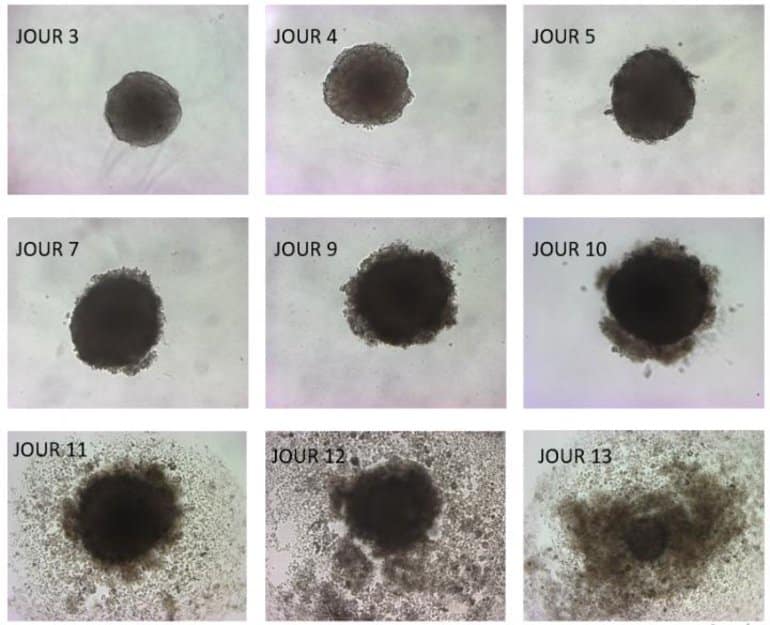
For their study, UCLouvain researchers used a 3D tumour cell culture system, called spheroids. In the presence of DHA, spheroids first grow and then implode. The team also administered a DHA-enriched diet to mice with tumours. The result: tumour development was significantly slowed compared to that in mice on a conventional diet.
This UCLouvain study shows the value of DHA in fighting cancer. “For an adult,” the UCLouvain researchers stated, “it’s recommended to consume at least 250 mg of DHA per day. But studies show that our diet provides on average only 50 to 100 mg per day. This is well below the minimum recommended intake.”
About this brain cancer research news
Source: UC Louvain
Contact: Isabelle Decoster – UC Louvain
Image: The image is credited to UC Louvain
Original Research: Closed access.
“Peroxidation of n-3 and n-6 polyunsaturated fatty acids in the acidic tumor environment leads to ferroptosis-mediated anticancer effects” by Feron et al. Cell Metabolism
Abstract
Peroxidation of n-3 and n-6 polyunsaturated fatty acids in the acidic tumor environment leads to ferroptosis-mediated anticancer effects
Tumor acidosis promotes disease progression through a stimulation of fatty acid (FA) metabolism in cancer cells. Instead of blocking the use of FAs by acidic cancer cells, we examined whether excess uptake of specific FAs could lead to antitumor effects. We found that n-3 but also remarkably n-6 polyunsaturated FA (PUFA) selectively induced ferroptosis in cancer cells under ambient acidosis.
Upon exceeding buffering capacity of triglyceride storage into lipid droplets, n-3 and n-6 PUFA peroxidation led to cytotoxic effects in proportion to the number of double bonds and even more so in the presence of diacylglycerol acyltransferase inhibitors (DGATi). Finally, an n-3 long-chain PUFA-rich diet significantly delayed mouse tumor growth when compared with a monounsaturated FA-rich diet, an effect further accentuated by administration of DGATi or ferroptosis inducers.
These data point out dietary PUFA as a selective adjuvant antitumor modality that may efficiently complement pharmacological approaches.


