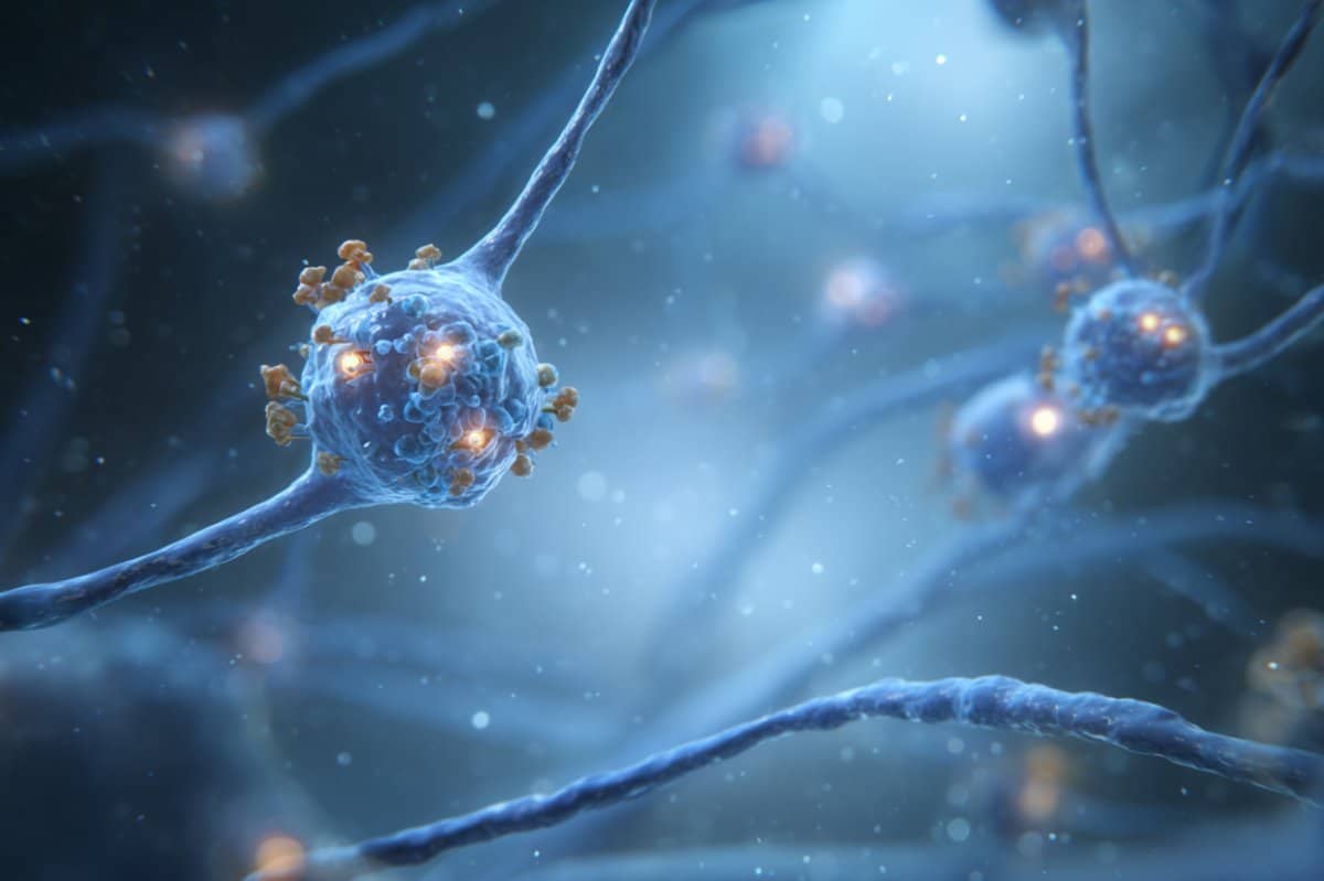
Summary: Scientists have mapped the molecular structure of glutamate receptors in the cerebellum for the first time using cryo-electron microscopy. These receptors are critical to how neurons in the cerebellum communicate, affecting movement, balance, and cognition.
By visualizing the receptors bound to proteins at synapses, researchers hope to inform future therapies that could restore function after injury or genetic disruption. While not immediately translatable to treatments, this foundational discovery offers a roadmap for repairing damaged brain circuits in motor and cognitive disorders.
Key Facts:
- First-Ever Visualization: Cryo-EM revealed the structure of cerebellar glutamate receptors at near-atomic resolution.
- Synaptic Precision Matters: The receptors are organized with exacting spatial precision to detect neurotransmitter signals.
- Therapeutic Potential: The findings lay groundwork for synapse-targeting therapies to address disorders involving movement and cognition.
Source: Oregon Health and Science University
For the first time, scientists using cryo-electron microscopy have discovered the structure and shape of key receptors connecting neurons in the brain’s cerebellum, which is located behind the brainstem and plays a critical role in functions such as coordinating movement, balance and cognition.
The research, published today in the journal Nature, provides new insight that could lead to the development of therapies to repair these structures when they are disrupted either by injury or genetic mutations affecting motor skills — sitting, standing, walking, running, and jumping — learning and memory.

The discovery by scientists at Oregon Health & Science University involves basic science research that won’t immediately lead to a new pill or treatment but is an example of an American commitment to medical research sustained over decades to advance human health.
Published in one of the world’s most prestigious scientific journals, the new research was supported by the National Institutes of Health and the Howard Hughes Medical Institute.
The study reveals the organization of a specific type of glutamate receptor — a chemical neurotransmitter that conveys signals between neurons and is considered the primary excitatory neurotransmitter in the brain — bound together with proteins clustered on synapses, or junctions, between neurons in the cerebellum.
“Synapses are crucial in all aspects of brain function, but the molecular structure has not been well understood in terms of how those pieces form together in a functional synapse,” said senior author Eric Gouaux, Ph.D., senior scientist with the OHSU Vollum Institute.
“It’s really critical to have receptors organized in exactly the right place so they can detect neurotransmitters released by an adjacent cell.”
Examining glutamate in the cerebellum
Using OHSU’s state-of-the-art cryo-electron microscopy, established as one of three national centers in 2018 and housed in the reinforced basement of a building on the university’s South Waterfront Campus, researchers examined the shape of a particular kind of glutamate receptor in the cerebellum of rodents at near-atomic scale.
“We know if there is an injury or genetic mutation in the cerebellum, it can lead to devastating disorders of balance, movement or cognition,” said co-author Laurence Trussell, Ph.D., professor of otolaryngology/head and neck surgery in the OHSU School of Medicine and a scientist in the Vollum Institute.
“This kind of glutamate receptor seems to be really important in how the cerebellum works. It’s entirely possible that developing drugs that target these receptors could improve its function.”
Gouaux, an investigator with the Howard Hughes Medical Institute and the Jennifer and Bernard Lacroute Endowed Chair in Neuroscience Research at OHSU, said the new discovery could have applications for new treatments.
“We’ve been interested in this question about synapse engineering and molecular insight that may one day help to repair damaged synapses,” he said. “This is a super exciting new direction with potential therapeutic applications.”
Lead author Chengli Fang, Ph.D., a postdoctoral researcher in the Gouaux lab, conducted almost all of the experiments reported in the publication.
In addition to Gouaux, Trussell and Fang, co-authors include Cathy J. Spangler, Ph.D., Jumi Park, Ph.D., of OHSU; and Natalie Sheldon of OHSU and the Howard Hughes Medical Institute.
Funding: The research reported in this publication was supported by the National Cancer Institute, the National Institute of Neurological Disorders and Stroke, and the National Institute of Deafness and Other Communication Disorders, all of the National Institutes of Health, award numbers K00CA253730, R01NS038631, R35NS116798 and R01DC004450.
The content is solely the responsibility of the authors and does not necessarily represent the official views of the NIH.
About this neuroscience research news
Author: Erik Robinson
Source: Oregon Health and Science University
Contact: Erik Robinson – Oregon Health and Science University
Image: The image is credited to Neuroscience News
Original Research: Closed Access.
“Gating and noelin clustering of native Ca2+-permeable AMPA receptors” by Eric Gouaux et al. Nature
Abstract
Gating and noelin clustering of native Ca2+-permeable AMPA receptors
AMPA-type ionotropic glutamate receptors (AMPARs) are integral to fast excitatory synaptic transmission and play vital roles in synaptic plasticity, motor coordination, learning, and memory.
While extensive structural studies have been conducted on recombinant AMPARs and native calcium impermeable (CI)-AMPARs alongside their auxiliary proteins, the molecular architecture of native calcium permeable (CP)-AMPARs has remained undefined.
To elucidate the subunit composition, physiological architecture, and gating mechanisms of CP-AMPARs, here we present the first visualization of these receptors, immunoaffinity purified from rat cerebella, and resolve their structures using cryo-electron microscopy (cryo-EM).
Our results indicate that the predominant assembly consists of GluA1 and GluA4 subunits, with the GluA4 subunit occupying the B and D positions, while auxiliary subunits, including TARPs, are located at the B′/D′ positions and CNIHs or TARPs at the A′/C′ positions.
Furthermore, we resolved the structure of the Noelin 1-GluA1/A4 complex, wherein Noelin 1 (Noe 1) specifically binds to the GluA4 subunit at the B and D positions. Notably, Noe 1 stabilizes the amino-terminal domain (ATD) layer without affecting receptor gating properties.
Noe 1 contributes to AMPAR function by forming dimeric-AMPAR assemblies that likely engage in extracellular networks, clustering receptors within synaptic environments and modulating receptor responsiveness to synaptic inputs.


