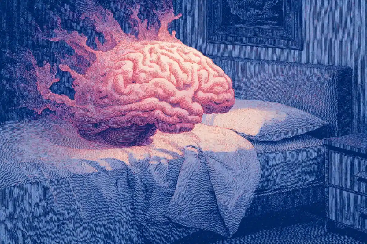
Summary: A collaborative neuroimaging study revealed that the duration of a depressive episode fundamentally alters how functional networks communicate inside the human brain. The research analyzed brain scans from patients with major depressive disorder who were not taking antidepressants, isolating the direct structural and functional footprints of the disease.
The study demonstrates that while short-term depression weakens the connection between executive control and introspective brain networks, long-term chronic depression completely flips this script, strengthening connectivity as symptoms worsen and trapping patients in a state of negative cognitive rumination.
Key Facts
- The Core Network Misalignment: Depression alters the coordinated dynamics between the Central Executive Network (CEN), which manages goal-oriented focus, and the Default Mode Network (DMN), which handles introspective self-reflection. This misalignment locks patients into repetitive, negative thoughts and impairs external concentration.
- The Chronicity Boundary: First author Tamires Zanão notes that the brain undergoes a structural evolution once depression crosses the chronic threshold of 24 months.
- The Precuneus Bridge: The precuneus region acts as a neural integration point or “bridge” between a person’s internal mental world and outward cognitive control.
- The Connection Reversal: In non-chronic patients, the functional connection between the executive network and the DMN’s precuneus decreases as symptoms worsen. In chronic, long-term patients, the opposite occurs: higher symptom severity correlates with stronger network connectivity.
- Gray Matter Changes: Higher severity of depressive symptoms directly associates with alterations in gray matter volume within the anterior cingulate cortex and the right dorsolateral prefrontal cortex.
- The Unmedicated Baseline: Because the 46-patient cohort was entirely unmedicated at the time of the brain scans, the findings cleanly isolate the neural architecture of depression itself, bypassing the brain-altering structural effects of antidepressants.
Source: FAPESP
Depression affects about 5.8% of the Brazilian population and presents a wide range of symptoms, intensities, and durations.
A study published in Scientific Reports involving patients with major depressive disorder demonstrated that the severity of symptoms, as measured by the Hamilton Depression Scale, and the length of time a person remains depressed (chronicity), are both associated with changes in brain function.
For the study, researchers from the University of São Paulo (USP) in Brazil and the University of Oxford in the United Kingdom analyzed brain images from 46 patients with major depression.
The results suggest that the duration of depressive episodes is associated with differences in functional brain connectivity. However, they do not allow for the differentiation or diagnosis of individual cases on their own.
“Major depression can alter brain function compared to people without the disorder. In this study, however, we identified that chronic patients [those with depression for more than 24 months] and non-chronic patients exhibit distinct patterns of connection between two important functional networks: the Central Executive Network [CEN], focused on executive control, and the Default Mode Network [DMN], associated with introspective thoughts and self-reflection,” says Tamires Zanão, a FAPESP fellowship recipient and first author of the study.
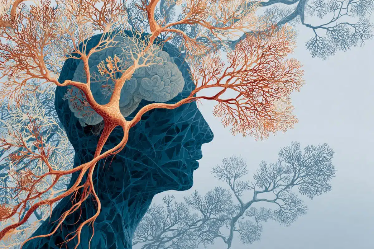
The researcher explains that the CEN and the DMN are two major brain systems with complementary functions. The CEN involves brain regions such as the dorsolateral prefrontal cortex and the parietal cortex. It is associated with the executive control required for goal-oriented tasks.
The DMN, on the other hand, is diffuse and includes areas such as the medial prefrontal cortex, the precuneus, and the hippocampus. It is related to internal processes such as self-reflection, autobiographical memory, and spontaneous thoughts. Due to its complexity, the DMN is often divided into subnetworks, such as those involving the precuneus.
“More detailed models of brain network organization suggest that the precuneus doesn’t act solely within the DMN, but also connects to subnetworks related to the CEN. For that reason, that region is considered a point of integration between different brain systems, functioning as a sort of ‘bridge’ between processes oriented toward the ‘internal world’ and cognitive control,” she says.
Typically, both networks exhibit coordinated dynamics with the participation of the salience network, which is involved in switching focus between the external environment and internal processes. In depression, however, these dynamics may be altered, which has been associated with symptoms such as rumination and difficulty concentrating.
“That misalignment between those networks may favor the predominance of introspective and self-referential thoughts, often with a negative bias. That helps explain why people with depression tend to get stuck in negative thoughts and have difficulty directing their attention to the environment when necessary,” Zanão explained to Agência FAPESP.
In the study, the researchers observed that the duration of depression appears to be associated with the dynamics of these two networks. In people with more recent episodes, the functional connection between the CEN and precuneal regions in the DMN decreases as symptoms worsen.
In contrast, patients with long-term depression exhibited an opposite pattern: the greater the severity, the stronger the connectivity between these networks.
Other studies using large population databases, such as the UK Biobank, have demonstrated a positive correlation between CEN activity and precuneus regions in the DMN in healthy individuals.
In the USP study conducted with the University of Oxford, although there was no direct comparison with healthy individuals, patients with non-chronic depression, especially those with fewer symptoms, exhibited connectivity patterns closer to the typical profile.
“The results are consistent with the hypothesis that changes in brain connectivity in depression may evolve over time. Previous research indicates that, in initial episodes, there may be a reduction in connectivity between certain networks, while in recurrent or more prolonged cases, changes in functional connectivity emerge,” Zanão explains.
Another finding from the study indicated an association between the severity of depressive symptoms and gray matter volume in two specific brain regions: the anterior cingulate cortex and the right dorsolateral prefrontal cortex. These regions have both been extensively linked to the disorder in previous studies.
“The anterior cingulate cortex, for example, has connections with areas involved in both emotional regulation and cognitive functions, playing an important role in integrating emotion and thought. Changes in that region have often been associated with the intensity of depressive symptoms.
“Although, in some contexts, greater gray matter volume is interpreted as indicative of better cognitive functioning, the results in the literature on depression are mixed.
“Previous studies have reported both reductions and increases in volume in those regions. Part of that discrepancy may be related to the use of antidepressants, as there’s evidence that medication can influence structural measures of the brain,” she states.
Because the current study included patients who were not taking antidepressants at the time of analysis, the authors suggest that the findings may more accurately reflect changes associated with depression itself rather than the effects of treatment.
Evidence from techniques such as tomography and transcranial magnetic stimulation suggests that depression may involve an imbalance in prefrontal cortex activity, with lower activity on the left side and higher activity on the right.
“According to that hypothesis, the left hemisphere would be more closely related to processing positive emotions, while the right hemisphere would be more involved with negative emotions.
“In this context, the finding of increased volume in the right dorsolateral prefrontal cortex observed in the study is consistent with that theoretical model, although its functional significance isn’t yet fully understood,” she says.
The researcher explains that these findings could aid in developing more personalized depression treatments in the future.
“Although guided by clinical evidence, the choice of treatment for depression still involves adjustments over time, as the response can vary from person to person. Studies like this one contribute to advancing our understanding of the disease, but more data is needed before this information can guide individualized clinical decisions,” she adds.
The brain imaging data from the 46 patients diagnosed with depression are part of a larger clinical trial coordinated by Professor André Brunoni from USP and currently at the University of Texas Southwestern Medical Center. Zanão analyzed the data for this study during his postdoctoral fellowship at the University of Oxford with support from FAPESP.
Funding: FAPESP also funded the research through projects 12/20911-5, 22/03266-0, and 23/13893-5.
Key Questions Answered:
A: It comes down to a literal physical misalignment between two massive systems in the brain. Your Central Executive Network (CEN) handles outward, goal-oriented tasks, while your Default Mode Network (DMN) handles inward self-reflection. Normally, a third system balances the two. In depression, this balance breaks, giving an absolute, negative bias to your inward thoughts and making it incredibly difficult to pull your attention back out to the real world.
A: This is the major discovery of the study: depression is a moving target that physically evolves over time. In the first 24 months of the illness, the connection between your executive brain and your inward-focused brain progressively weakens as you feel worse. But if depression becomes chronic (lasting over two years), the brain rewires itself. It enters an opposite pattern where severe symptoms actually lock those networks tighter together, cementing the depressive loop.
A: While these neuroimaging results are monumental for understanding the group-level biology of depression, the connectivity maps are not yet distinct enough to reliably diagnose a single individual case on its own. Everyone’s baseline brain architecture is unique. However, uncovering these independent, time-dependent network changes gives drug developers and psychiatrists a brilliant roadmap to design custom therapies tailored to how long a patient has been suffering.
Editorial Notes:
- This article was edited by a Neuroscience News editor.
- Journal paper reviewed in full.
- Additional context added by our staff.
About this depression and neuroscience research news
Author: Heloisa Reinert
Source: FAPESP
Contact: Heloisa Reinert – FAPESP
Image: The image is credited to Neuroscience News
Original Research: Open access.
“Chronicity moderates the impact of severity on central executive-default mode network functional interactions in depression” by Tamires Zanao, Piergiorgio Salvan, Lais B. Razza, Pedro Henrique Rodrigues da Silva, Andre R. Brunoni & Jacinta O’Shea. Scientific Reports
DOI:10.1038/s41598-026-40364-2
Abstract
Chronicity moderates the impact of severity on central executive-default mode network functional interactions in depression
Neuroimaging has revealed that major depression is underpinned by dysfunctional brain networks, with symptom variability stemming from altered interactions within and between brain regions. While the effect of depression severity is well-studied, the effect of depression duration (chronicity) is relatively neglected, despite its clinical significance.
This study examined how severity, chronicity, and their interaction affect brain network connectivity and grey matter volume. Forty-six patients (31 females, mean age 40.5) were assessed using whole-brain network modeling and voxel-based morphometry (VBM). Severity was measured via the Hamilton Depression Rating Scale, and chronicity was defined as an episode lasting over 24 months.
The key finding was that chronicity moderated the impact of severity on functional connectivity between the Central Executive Network (CEN) and the precuneus (part of the Default Mode Network, DMN). Chronic versus non-chronic patients showed opposite patterns.
Non-chronic patients showed stronger CEN-Default Mode Precuneus connectivity at low severity and weaker at high severity; chronic patients showed the reverse.
This study reveals a novel impact of chronicity on CEN-DMN interactions, a neglected moderator of brain-symptom severity correlations in depression.