

Summary: Axonal swelling in the Purkinje cells of mice had no detrimental impact on firing rate or the speed at which axons transmit signals. At peak firing rate, axons with swellings were less likely to fail than those without.
Source: McGill University
Researchers at McGill University have shown that a brain cell structure previously thought to be pathological in fact enhances cells’ ability to transmit information and correlates with better learning on certain tasks.
In a study published in Nature Communications, the team investigated swellings that occur in the axons of Purkinje cells in the cerebellum. In results that contradict established expectations, they found that axons with swellings did a better job of conducting electrical signals than those without.
“These are the kind of results where you really scratch your head and think, ‘Let’s check that again’,” says senior author Alanna Watt, an Associate Professor in the Department of Biology. “We really thought when we started this that we were going to characterize and measure how an axon fails – and that’s not at all what we saw.”
Experimental data confounds expectations
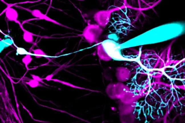
Swellings in axons – the long, slender fibres through which nerve cells transmit information to other cells – are observed in normal development and in disease. Increased numbers of axonal swellings are seen in various neurodegenerative disorders, which has led scientists to believe swellings have a negative impact on axon function. While computer modelling lends some support to this view, the McGill research team was the first to test the theory with measurements on actual nerve cells.
Using a technically challenging combination of two-photon microscopy and electrophysiology to measure electrical activity simultaneously at different locations within cells, the researchers demonstrated that the presence of axonal swellings in mouse Purkinje cells had no detrimental impact on the rate at which those cells produced signals (the firing rate) or on the speed at which axons transmitted the signals. Remarkably, the experiments also showed that at peak firing rates axons with swellings were less likely to fail than those without.
Role in neurodegenerative disease called into question
In a surprising twist, the McGill researchers found that they were able to stimulate the formation of axonal swellings by introducing a pharmaceutical that blocked the transmission of electrical signals in nerve cells, especially in the axons. Seeing axonal swellings form within a matter of hours when the axon of a nerve cell had been compromised in this way challenges previous assumptions about the role of axonal swellings in neurodegenerative disorders. As Watt explains, the discovery leaves room for the possibility that swellings represent a self-repair mechanism rather than a deterioration caused by disease.
“Transmitting information by way of electrical signals is the most important thing that an axon does,” she says. “If it starts to fail at that job, it makes sense that there’s a mechanism that tries to prevent that.”
Behavioural analysis confirms swellings’ positive impact
In addition to their investigations at the cellular level, the researchers sought to determine the impact of axonal swellings on overall brain function. The team used three tests designed to assess motor learning and coordination, which are among the cerebellum’s most important roles.
Supported by modeling to account for the natural variation between individual learning abilities, the results revealed a positive correlation between the abundance of axonal swellings in cerebellar Purkinje cells and motor learning abilities.
“We think the link is probably an indirect one,” Watt explains. “The learning is probably happening somewhere else, but the information is being relayed more reliably and therefore we see an improvement.”
About this neuroscience research news
Source: McGill University
Contact: Frédérique Mazerolle – McGill University
Image: The image is credited to Kim Gruver, Watt Lab.
Original Research: Open access.
“Purkinje cell axonal swellings enhance action potential fidelity and cerebellar function” by Daneck Lang-Ouellette et al. Nature Communications
Abstract
Purkinje cell axonal swellings enhance action potential fidelity and cerebellar function
Axonal plasticity allows neurons to control their output, which critically determines the flow of information in the brain. Axon diameter can be regulated by activity, yet how morphological changes in an axon impact its function remains poorly understood. Axonal swellings have been found on Purkinje cell axons in the cerebellum both in healthy development and in neurodegenerative diseases, and computational models predicts that axonal swellings impair axonal function.
Here we report that in young Purkinje cells, axons with swellings propagated action potentials with higher fidelity than those without, and that axonal swellings form when axonal failures are high.
Furthermore, we observed that healthy young adult mice with more axonal swellings learn better on cerebellar-related tasks than mice with fewer swellings.
Our findings suggest that axonal swellings underlie a form of axonal plasticity that optimizes the fidelity of action potential propagation in axons, resulting in enhanced learning.


