Summary: Astrocytes may play a significant role in information processing and memory, a new study reports.
Source: OIST
The way people experience the world occurs due to complex and intricate interactions between neurons in the brain.
Now, a study, published 9th February 2022 in Science Advances, suggests that astrocytes—star-shaped, non-neuronal cells in the brain—might also play an important role in processing information, and perhaps even memory.
Using advanced imaging and analysis techniques, researchers from the Okinawa Institute of Science and Technology Graduate University (OIST) recorded signaling within single astrocytes at a previously unseen level of detail and speed in the brains of awake mice.
Their findings, including ultra-fast signals on par with those seen in neurons and patterns of signaling activity that correspond to different behaviors, suggest that astrocytes may play a crucial role in many functions of our brain, including how we think, move, and learn.
“If these implications are true, it will fundamentally transform how we think about neuroscience, and the way the brain works,” said first author Dr. Leonidas Georgiou, a former Ph.D. student in the Optical Neuroimaging Unit at OIST.
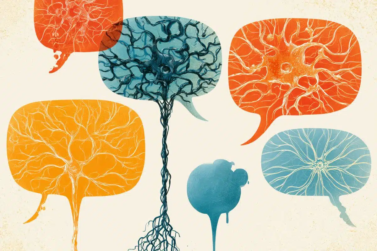
When we picture our brain, we typically imagine a messy tangle of long, wire-like neurons that send electrical signals to each other across different regions of the brain. But neurons only make up half the cells in our brain. Crammed into all the remaining space between the jumble of neurons are many other types of brain cells, including astrocytes.
“Compared to neurons, astrocytes have received very little attention. It was thought that astrocytes are just helper cells, supplying the neurons with nutrients and removing their waste,” said Professor Bernd Kuhn, senior author and head of the Optical Neuroimaging Unit.
But in recent years, there’s been increasing amounts of evidence that astrocytes can listen to chemical messages sent between neurons at synapses, and can respond with their own signals, providing an extra layer of complexity to how our brain receives and responds to information.
Still, the previously detected signals in astrocytes were about ten times slower than signals seen in neurons, with scientists therefore believing the cells were too slow for information processing.
However, by developing a new toolkit that allows the study of astrocyte activity in awake mice with unprecedented detail, the researchers at OIST showed for the first time that astrocytes generate signals in vivo which are as fast as that of neurons, lasting fewer than 300 milliseconds.
Their toolkit relied on a new discovery: that a virus regularly used for gene therapy could “jump” from neurons to connected astrocytes. The scientists used an adeno-associated virus that contained a gene that makes infected cells fluoresce. The fluorescence increases in intensity in the presence of calcium—an important indicator of signal activity within living cells.
Once labeled, the research team were able to use a powerful, homebuilt microscope to pinpoint and image a single astrocyte, over multiple days for up to an hour at a time, while the mouse was awake and moving.
The scientists then used an advanced computer program to analyze the recorded images, allowing them to detect the never-before-seen ultra-fast flashes of calcium signals, and evaluate signal patterns in an unbiased way.
They found that sensory stimulation, by tickling the whiskers, resulted in very little calcium signaling, while certain behaviors, like running or walking, resulted in high levels of activity.
The scientists also realized that there were certain areas in the astrocyte, or hotspots, where levels of activity were higher.

“These hotspot maps are like fingerprints—for a specific behavior, they are stable over time, remaining the same over a period of days, and unique to each astrocyte,” said Dr. Georgiou.
Even more surprisingly, the team noticed that different behaviors corresponded to unique hotspot patterns.
“So when the mouse is resting, you see one pattern. And then when the mouse is running, you see a different pattern,” said Prof. Kuhn.
One hypothesis suggested by Prof. Kuhn is that these hotspot maps could represent memory engrams—a pattern that represents a specific behavior or a memory. Different neuron networks are active during specific behaviors or when learning and recalling information, which could also change the activity of nearby astrocytes. Memory engrams are still theoretical, and highly controversial, he acknowledged.
“We still don’t know how memories are stored in a brain, but it’s incredible to think that it could involve astrocytes,” he said. “It’s likely too good to be true, but it’s an exciting hypothesis to follow up on.”
About this neuroscience research news
Author: Press Office
Source: OIST
Contact: Press Office – OIST
Image: The image is credited to OIST
Original Research: Open access.
“Ca+ activity maps of astrocytes tagged by axoastrocytic AAV transfer” by Leonidas Georgiou et al. Science Advances
Abstract
Ca+ activity maps of astrocytes tagged by axoastrocytic AAV transfer
Astrocytes exhibit localized Ca2+ microdomain (MD) activity thought to be actively involved in information processing in the brain. However, functional organization of Ca2+ MDs in space and time in relationship to behavior and neuronal activity is poorly understood.
Here, we first show that adeno-associated virus (AAV) particles transfer anterogradely from axons to astrocytes. Then, we use this axoastrocytic AAV transfer to express genetically encoded Ca2+ indicators at high-contrast circuit specifically. In combination with two-photon microscopy and unbiased, event-based analysis, we investigated cortical astrocytes embedded in the vibrissal thalamocortical circuit.
We found a wide range of Ca2+ MD signals, some of which were ultrafast (≤300 ms). Frequency and size of signals were extensively increased by locomotion but only subtly with sensory stimulation. The overlay of these signals resulted in behavior-dependent maps with characteristic Ca2+ activity hotspots, maybe representing memory engrams.
These functional subdomains are stable over days, suggesting subcellular specialization.