Summary: FMR1, a gene mutation that causes Fragile X syndrome is also implicated in premature ovarian failure, resulting in infertility in females. The mutation alters neurons that regulate reproduction in the brain and ovaries.
Source: UCR
A University of California, Riverside, study has identified the biological underpinnings of a reproductive disorder caused by the mutation of a gene. This gene mutation also causes Fragile X Syndrome, a leading genetic cause of intellectual impairment and autism.
The researchers found mutations of the Fragile X messenger ribonucleoprotein 1 gene, or FMR1, contribute to premature ovarian failure, or POF, due to changes in neurons that regulate reproduction in the brain and ovaries. The mutation has been associated with early infertility, due to a 25-fold increased risk of POF, but the reasons were unclear.
POF is the most severe form of premature ovarian aging, which affects about 10% of women and is characterized by an early depletion of ovarian follicles and early menopause. With women postponing reproduction, the chances of infertility increase, including due to FMR1 mutation.
“In the last two or three decades, the median age of first-time mothers in the U.S. and Europe has steadily increased,” said Djurdjica Coss, a professor of biomedical sciences in the UCR School of Medicine who led the research team.
“Moreover, premature menopause causes not only early infertility, but also increased risk of cardiovascular disease and osteoporosis. It’s important, therefore, to understand the reasons behind these reproductive disorders and eventually find treatments. Such research can also help better advise women at risk on when to have a child and how to monitor their health outcomes.”
According to the Centers for Disease Control and Prevention, 19% of heterosexual couples in the U.S. experience infertility and need assisted reproductive technology, which can be too costly for many couples.
Coss explained that previous studies concerning the FMR1-mediated reproductive disorders analyzed them exclusively from an endocrine perspective, meaning they studied the changes in hormone levels and how endocrine cells functioned in the ovaries that produce them.
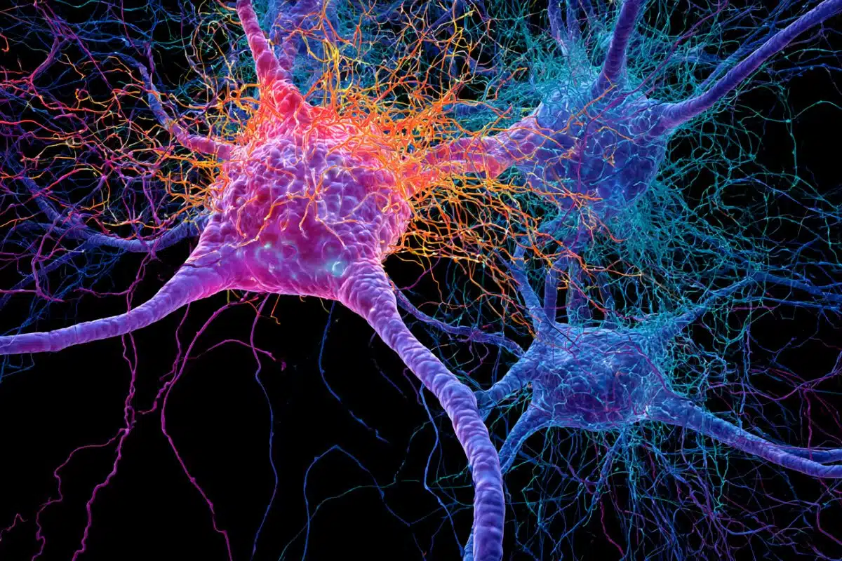
“We took a different approach,” Coss said. “Since the FMR1 gene is highly abundant in neurons, we postulated that neurons that regulate reproduction are affected by the FMR1 mutation, which in turn causes increases in hormone levels.
“Indeed, we found higher stimulation of neurons in the hypothalamus that regulate reproduction as well as more neurons in the ovaries that contribute to ovarian hormone synthesis.”
To do the research, Coss and her team used transgenic mice that lack the FMR1 gene to emulate the condition in people with a mutation in this gene. They first determined that this mouse model mimics what is observed in women with a FMR1 mutation. They then compared the reproduction-regulating neurons in the ovaries and the brain between these mice and their normal counterparts.
They found the changes in function of these neurons led to a more rapid secretion of hormones in young transgenic female mice that later stopped reproducing early. Next, they removed the ovaries from these mice to determine the effect of the FMR1 mutation on just the neurons in the brain.

“This allowed us to determine that these neurons in the brain, called gonadotropin-releasing hormone neurons, show changes in connectivity that affect how they function,” Coss said. “The increased number of synapses cause them to be faster and have more pulses of hormone secretion.”
Her team also determined that neurons “innervating” the ovaries — supplying the ovaries with nerves — were more abundant in the transgenic mice than in their normal counterparts.
“We think the increases we see in ovarian hormone levels are due to increases in ovarian innervation rather than increases in hormone-producing cells,” Coss said. “The endocrine perspective supports the latter.”
Next, Coss and her team plan to investigate if the effects of FMR1 mutation can be alleviated by partially inhibiting neurons in the ovaries.
“We anticipate this may normalize ovarian hormone levels, possibly allowing for a normal reproductive lifespan,” Coss said.
Coss was joined in the study by Pedro A. Villa, Nancy M. Lainez, Carrie R. Jonak, Sarah C. Berlin, and Iryna M. Ethell.
Funding: The study, published in the journal Frontiers in Endocrinology, was supported by a grant from the Eunice Kennedy Shriver National Institute of Child Health and Human Development of the National Institutes of Health.
About this ASD and genetics research news
Author: Iqbal Pittalwala
Source: UCR
Contact: Iqbal Pittalwala – UCR
Image: The image is in the public domain
Original Research: Open access.
“Altered GnRH neuron and ovarian innervation characterize reproductive dysfunction linked to the Fragile X messenger ribonucleoprotein (Fmr1) gene mutation” by Djurdjica Coss et al. Frontiers in Endocrinology
Abstract
Altered GnRH neuron and ovarian innervation characterize reproductive dysfunction linked to the Fragile X messenger ribonucleoprotein (Fmr1) gene mutation
Introduction: Mutations in the Fragile X Messenger Ribonucleoprotein 1 (FMR1) gene cause Fragile X Syndrome, the most common monogenic cause of intellectual disability. Mutations of FMR1 are also associated with reproductive disorders, such as early cessation of reproductive function in females. While progress has been made in understanding the mechanisms of mental impairment, the causes of reproductive disorders are not clear. FMR1-associated reproductive disorders were studied exclusively from the endocrine perspective, while the FMR1 role in neurons that control reproduction was not addressed.
Results: Here, we demonstrate that similar to women with FMR1 mutations, female Fmr1 null mice stop reproducing early. However, young null females display larger litters, more corpora lutea in the ovaries, increased inhibin, progesterone, testosterone, and gonadotropin hormones in the circulation. Ovariectomy reveals both hypothalamic and ovarian contribution to elevated gonadotropins. Altered mRNA and protein levels of several synaptic molecules in the hypothalamus are identified, indicating reasons for hypothalamic dysregulation. Increased vascularization of corpora lutea, higher sympathetic innervation of growing follicles in the ovaries of Fmr1 nulls, and higher numbers of synaptic GABAA receptors in GnRH neurons, which are excitatory for GnRH neurons, contribute to increased FSH and LH, respectively. Unmodified and ovariectomized Fmr1 nulls have increased LH pulse frequency, suggesting that Fmr1 nulls exhibit hyperactive GnRH neurons, regardless of the ovarian feedback.
Conclusion: These results reveal Fmr1 function in the regulation of GnRH neuron secretion, and point to the role of GnRH neurons, in addition to the ovarian innervation, in the etiology of Fmr1-mediated reproductive disorders.