Summary: A new study reveals the majority of V1 neurons project to higher visual areas in a non-random manner.
Source: Sainsbury Wellcome Center.
An international collaboration of neuroscientists have today published a paper in Nature demonstrating the breadth of neural communication in visual cortex using a combination of methods for tracing the projections of individual neurons across the brain.
In classical models of the visual system, information flows from ‘primary’ visual cortex (V1) to more specialized, downstream areas that focus for example on image movement or image form. However, the details of how individual cells carry this information are not understood.
Professor Tom Mrsic-Flogel, one of the senior authors of the paper and project leader at Biozentrum, University of Basel and Director of the Sainsbury Wellcome Centre commented:
“Understanding the fine-scale anatomy by which individual neurons distribute signals to their targets is a crucial step for forging the relationship between neuronal structure and function.”
Up until now, it had remained unclear as to whether information transfer from primary visual cortex was largely “one neuron – one target area”, or if individual neurons distributed their signals across multiple downstream areas.
While the research, conducted by neuroscientists from the Sainsbury Wellcome Centre for Neural Circuits and Behaviour, in conjunction with Cold Spring Harbor and Biozentrum, confirmed the existence of dedicated projections to certain cortical areas; the scientists found that these were the exception and that the majority of primary visual cortex neurons broadcast information to multiple targets.
Justus M. Kebschull, one of the first authors on the paper at Cold Spring Harbor commented:
“Our findings reveal that individual neurons in the visual cortex project to several targets in the neocortex. This means that their signals are distributed widely and that individual neurons contribute to multiple parallel computations across the neocortex.”
In the Nature paper, the team outline the two complementary methods they used to map the projection patterns. Firstly, they used whole-brain fluorescence-based axonal tracing by labelling neurons in the right visual cortex of each mouse with GFP and then imaging axonal projections by whole-brain serial two-photon tomography.
The Allen Mouse Brain Reference Atlas was then used to identify the areas in which axonal terminations were observed. The mouse primary visual cortex (V1) neurons were found to have a high degree of projectional diversity and most of the individual layer 2/3 neurons were found to distribute information to multiple areas rather than projecting to a single target. Such neurons were termed ‘broadcasting neurons’.
Secondly, the researchers used high-throughput DNA sequencing of genetically barcoded neurons (MAPseq) to determine whether the broadcasting neurons were targeting cortical areas at random, or whether they preferentially target, or avoid, specific subsets of areas thereby indicating a higher-order structure.
Thousands of individual V1 neurons were uniquely labelled with random RNA sequences, in essence barcodes. Each labelled neuron then transports the barcode into its own axonal processes where they can be read out by high throughout sequencing of a dissected target area to determine the projection targets of that specific neuron to higher visual areas.
Professor Anthony Zador, another senior author and project leader at Cold Spring Harbor Laboratory, explains the revolutionary technique:
“The RNA sequences, or ‘barcodes’, that we deliver to individual neurons are unmistakably unique and this enables us to determine if individual neurons, as opposed to entire regions, are tailored to specific targets.”
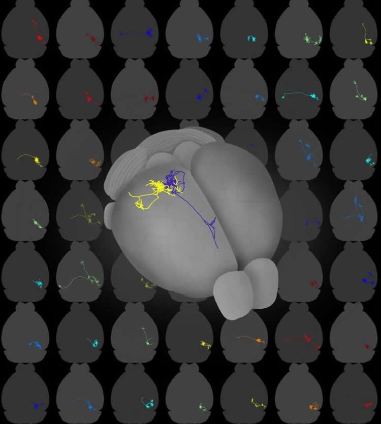
This technique revealed that the majority of V1 neurons project to higher visual areas in a non-random manner. Six projection motifs were identified that reflect several sub-classes of projection neurons for divergent information transfer from V1 to higher visual areas.
The researchers state that the results of the study “suggest a functional specialization of subpopulations of projection cells beyond ‘one neuron – one target area’ mapping.
“The next piece of the puzzle will be to understand what each of these projection motifs does for visual processing and perception and how these long-range connectivity patterns are established during development,” Professor Mrsic-Flogel concluded.
Funding: This research was supported by U.S. National Institutes of Health; Brain Research Foundation; IARPA; Simons Foundation; Paul Allen Distinguished Investigator Award; Boehringer Ingelheim Fonds; Genentech Foundation; European Research Council, and Swiss National Science Foundation.
Source: April Cashin-Garbutt – Sainsbury Wellcome Center
Publisher: Organized by NeuroscienceNews.com.
Image Source: NeuroscienceNews.com image is credited to Sainsbury Wellcome Centre.
Original Research: Abstract for “The logic of single-cell projections from visual cortex” by Yunyun Han, Justus M. Kebschull, Robert A. A. Campbell, Devon Cowan, Fabia Imhof, Anthony M. Zador & Thomas D. Mrsic-Flogel in Nature. Published online March 28 2018.
doi:10.1038/nature26159
[cbtabs][cbtab title=”MLA”]Sainsbury Wellcome Center “Brain Wide Tracing of Single Neurons Reveals Breadth of Information Transfer From Visual Cortex.” NeuroscienceNews. NeuroscienceNews, 28 March 2018.
<https://neurosciencenews.com/visual-cortex-neuron-tracing-8704/>.[/cbtab][cbtab title=”APA”]Sainsbury Wellcome Center (2018, March 28). Brain Wide Tracing of Single Neurons Reveals Breadth of Information Transfer From Visual Cortex. NeuroscienceNews. Retrieved March 28, 2018 from https://neurosciencenews.com/visual-cortex-neuron-tracing-8704/[/cbtab][cbtab title=”Chicago”]Sainsbury Wellcome Center “Brain Wide Tracing of Single Neurons Reveals Breadth of Information Transfer From Visual Cortex.” https://neurosciencenews.com/visual-cortex-neuron-tracing-8704/ (accessed March 28, 2018).[/cbtab][/cbtabs]
Abstract
The logic of single-cell projections from visual cortex
Neocortical areas communicate through extensive axonal projections, but the logic of information transfer remains poorly understood, because the projections of individual neurons have not been systematically characterized. It is not known whether individual neurons send projections only to single cortical areas or distribute signals across multiple targets. Here we determine the projection patterns of 591 individual neurons in the mouse primary visual cortex using whole-brain fluorescence-based axonal tracing and high-throughput DNA sequencing of genetically barcoded neurons (MAPseq). Projections were highly diverse and divergent, collectively targeting at least 18 cortical and subcortical areas. Most neurons targeted multiple cortical areas, often in non-random combinations, suggesting that sub-classes of intracortical projection neurons exist. Our results indicate that the dominant mode of intracortical information transfer is not based on ‘one neuron–one target area’ mapping. Instead, signals carried by individual cortical neurons are shared across subsets of target areas, and thus concurrently contribute to multiple functional pathways.