Summary: Cerebral blood flow is reduced in the Broca’s area of people who stutter, researchers report. Additionally, the more severely a person stutters, the less blood flows to this area of the brain.
Source: Children’s Hospital of Los Angeles.
A study led by researchers at Children’s Hospital Los Angeles demonstrates what lead investigator Bradley Peterson, MD, calls “a critical mass of evidence” of a common underlying lifelong vulnerability in both children and adults who stutter. They discovered that regional cerebral blood flow is reduced in the Broca’s area – the region in the frontal lobe of the brain linked to speech production – in persons who stutter. More severe stuttering is associated with even greater reductions in blood flow to this region.
In addition, a greater abnormality of cerebral blood flow in the posterior language loop, associated with processing words that we hear, correlates with more severe stuttering. This finding suggests that a common pathophysiology throughout the neural “language” loop that connects the frontal and posterior temporal lobe likely contributes to stuttering severity.
Peterson, who is director of the Institute for the Developing Mind at CHLA and a professor of the Keck School of Medicine at the University of Southern California, says that such a study of resting blood flow, or perfusion, has never before been conducted in persons who stutter. His team also recently published a study using proton magnetic resonance spectroscopy to look at brain regions in both adults and children who stutter. Those findings demonstrated links between stuttering and changes in the brain circuits that control speech production, as well as those supporting attention and emotion. The present blood flow study adds significantly to the findings from that prior study and furthermore suggests that disturbances in the speech processing areas of the brain are likely of central importance as a cause of stuttering.
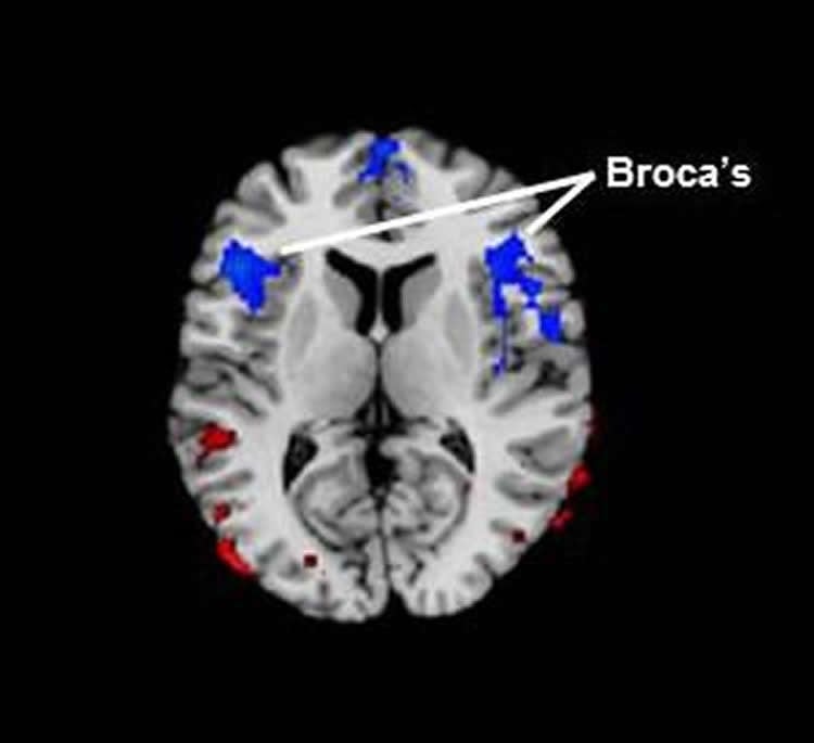
According to Peterson, the new study – published on December 30 in the journal Human Brain Mapping – provides scientists with a completely different window into the brain. The researchers were able to zero in on the Broca’s area as well as related brain circuitry specifically linked to speech, using regional cerebral blood flow as a measure of brain activity, since blood flow is typically coupled with neural activity.
“When other portions of the brain circuit related to speech were also affected according to our blood flow measurements, we saw more severe stuttering in both children and adults,” said first author Jay Desai, MD, a clinical neurologist at CHLA. “Blood flow was inversely correlated to the degree of stuttering – the more severe the stuttering, the less blood flow to this part of the brain,” said Desai, adding that the study results were “quite striking.”
Additional contributors to the study include Ravi Bansal, Children’s Hospital Los Angeles and the Keck School of Medicine of USC; Yuankai Huo and Zhishun Wang, Columbia University; Steven C. R. Williams, David Lythgoe and Fernando O. Zelaya, King’s College, London, UK.
Funding: This work was supported in part by Children’s Hospital Los Angeles, the National Institute of Mental Health grant K0274677, the Milhiser family fund and the Murphy endowment at Columbia University.
Source: Ellin Kavanagh – Children’s Hospital of Los Angeles
Image Source: NeuroscienceNews.com image is credited to Children’s Hospital Los Angeles.
Original Research: Abstract for “Reduced perfusion in Broca’s area in developmental stuttering” byJay Desai, Yuankai Huo, Zhishun Wang, Ravi Bansal, Steven C. R. Williams, David Lythgoe, Fernando O. Zelaya and Bradley S. Peterson in Human Brain Mapping. Published online December 30 2016 doi:10.1002/hbm.23487
[cbtabs][cbtab title=”MLA”]Children’s Hospital of Los Angeles “Stuttering Linked to Reduced Blood Flow in Brain Area Associated With Language.” NeuroscienceNews. NeuroscienceNews, 4 January 2017.
<https://neurosciencenews.com/stuttering-blood-flow-brocas-5853/>.[/cbtab][cbtab title=”APA”]Children’s Hospital of Los Angeles (2017, January 4). Stuttering Linked to Reduced Blood Flow in Brain Area Associated With Language. NeuroscienceNew. Retrieved January 4, 2017 from https://neurosciencenews.com/stuttering-blood-flow-brocas-5853/[/cbtab][cbtab title=”Chicago”]Children’s Hospital of Los Angeles “Stuttering Linked to Reduced Blood Flow in Brain Area Associated With Language.” https://neurosciencenews.com/stuttering-blood-flow-brocas-5853/ (accessed January 4, 2017).[/cbtab][/cbtabs]
Abstract
Reduced perfusion in Broca’s area in developmental stuttering
Objective
To study resting cerebral blood flow in children and adults with developmental stuttering.
Methods
We acquired pulsed arterial spin labeling magnetic resonance imaging data in 26 participants with stuttering and 36 healthy, fluent controls. While covarying for age, sex, and IQ, we compared perfusion values voxel-wise across diagnostic groups and assessed correlations of perfusion with stuttering severity within the stuttering group and with measures of motor speed in both groups.
Results
We detected lower regional Cerebral Blood Flow (rCBF) at rest in the stuttering group compared with healthy controls in Broca’s area bilaterally and the superior frontal gyrus. rCBF values in Broca’s area bilaterally correlated inversely with the severity of stuttering and extended posteriorly into other portions of the language loop. We also found increased rCBF in cerebellar nuclei and parietal cortex in the stuttering group compared with healthy controls. Findings were unchanged in child-only analyses and when excluding participants with comorbid illnesses or those taking medication.
Conclusions
rCBF is reduced in Broca’s region in persons who stutter. More severe stuttering is associated with even greater reductions in rCBF to Broca’s region, additive to the underlying putative trait reduction in rCBF relative to control values. Moreover, a greater abnormality in rCBF in the posterior language loop is associated with more severe symptoms, suggesting that a common pathophysiology throughout the language loop likely contributes to stuttering severity.
“Reduced perfusion in Broca’s area in developmental stuttering” byJay Desai, Yuankai Huo, Zhishun Wang, Ravi Bansal, Steven C. R. Williams, David Lythgoe, Fernando O. Zelaya and Bradley S. Peterson in Human Brain Mapping. Published online December 30 2016 doi:10.1002/hbm.23487