A method known as navigated transcranial magnetic stimulation (nTMS) has been gaining importance in neurosurgery for some time now. Among other applications, it is used to map brain tumors before an operation and to test whether important regions of the brain, for example motor and language areas, are affected. Doctors at the Technische Universität München (TUM) have now shown that preoperative nTMS analysis of motor areas improves the prognosis of patients with malignant brain tumors.
With the help of nTMS, it is possible to identify what areas of the brain control motor or language function to an accuracy of four millimeters. “That’s particularly important, as it enables the removal of tumors from patients without affecting functional areas while at the same time removing as much of the malignant tissue as possible,” explains Dr. Sandro Krieg, working group leader at the Department of Neurosurgery of TUM University Hospital Klinikum rechts der Isar and head of the study. Mapping must be performed separately for each patient, as tumors can displace important brain areas from their original sites.
Map for important motor areas
To determine motor areas with the nTMS technique, the physician scans points at defined positions on the patient’s head using a coil. The coil induces brief painless electrical pulses in the brain, which stimulate brain neurons at those positions. If the pulses at a given position point activate neurons that trigger muscle movements, the scientists are able to measure the muscle activities with the help of electrodes fastened to the patient’s arms and legs. That position is then regarded as an essential location for motor activity.
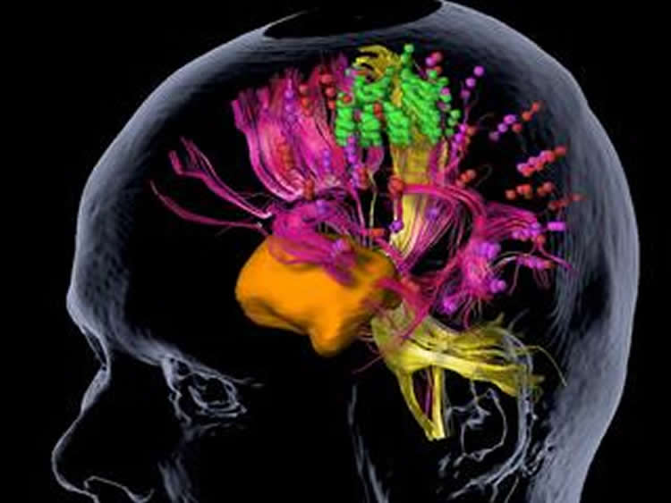
Up to 150 individual position points per patient can be analyzed and mapped. “In this way, we can draw up a map of important motor areas and nerve pathways in the vicinity of the tumor. During the operation, this data is a good guide as to where we can and cannot cut,” explains Krieg. The method has been used at Klinikum rechts der Isar since 2010.
nTMS analysis improves prognosis
In their recent study, Sandro Krieg and his team scanned and mapped motor areas in patients with high-grade gliomas (HGG), which are usually associated with a poor prognosis. They compared two groups: 70 subjects who underwent preoperative nTMS analysis and 70 patients who underwent tumor surgery before nTMS became a standard procedure in the hospital.
The results of the study show the advantage of nTMS mapping for patients compared to the control group: smaller openings in the skull had to be made in patients who underwent nTMS tumor analysis, and residual tumor tissue was left behind less frequently. In addition, their average hospitalization period was two days shorter. Because the general state of health of the nTMS-treated patients was also better, an increased number of them were subsequently able to receive radiotherapy. Above all, those patients survived several months longer than the control group.
“Of course, we need to confirm the findings in larger patient groups, but one important thing is clear: preoperative tumor mapping positively influences many aspects of the procedure,” Krieg says about the results, adding: “Some tumors that are otherwise thought to be inoperable can be removed by this method.”
Refining protocols for nTMS
The scientists now aim to improve standard protocols for nTMS and, for example, the mapping of language areas. In a recent study, they showed that an object-naming test is the best method for analyzing language centers. “Language areas can already be mapped with nTMS, but they’re much more complex than motor areas. We want to create higher standards in order to help patients with tumors in these regions as effectively as possible,” the scientist says.
Funding: Funding was provided by the Max Planck Society.
Source: Vera Siegler – TUM
Image Source: The image is credited to Sandro Krieg / TUM
Original Research: 1) Full open access research for “Changing the clinical course of glioma patients by preoperative motor mapping with navigated transcranial magnetic brain stimulation” by S. M. Krieg, N. Sollman, T. Obermüller, J. Sabih, L. Bulubas, C. Negwer, T. Moser, D. Droese, T. Boeckh-Behrens, F. Ringel, and B. Meyer in BMC Cancer. Published online April 8 2015 doi:10.1186/s12885-015-1258-1
2) Full open access research for “Task Type Affects Location of Language-Positive Cortical Regions by Repetitive Navigated Transcranial Magnetic Stimulation Mapping” by T. Hauck, N. Tanigawa, M. Probst, A. Wohlschlaeger, S. Ille, N. Sollmann, S. Maurer, C. Zimmer, F. Ringel, B. Meyer, S. M. Krieg in PLOS ONE. Published online April 30 2015 doi:10.1371/journal.pone.0125298
Abstract
Changing the clinical course of glioma patients by preoperative motor mapping with navigated transcranial magnetic brain stimulation
Background
Mapping of the motor cortex by navigated transcranial magnetic stimulation (nTMS) can be used for preoperative planning in brain tumor patients. Just recently, it has been proven to actually change outcomes by increasing the rate of gross total resection (GTR) and by reducing the surgery-related rate of paresis significantly in cohorts of patients suffering from different entities of intracranial lesions. Yet, we also need data that shows whether these changes also lead to a changed clinical course, and can also be achieved specifically in high-grade glioma (HGG) patients.
Methods
We prospectively enrolled 70 patients with supratentorial motor eloquently located HGG undergoing preoperative nTMS (2010–2014) and matched these patients with 70 HGG patients who did not undergo preoperative nTMS (2007–2010).
Results
On average, the overall size of the craniotomy was significantly smaller for nTMS patients when compared to the non-nTMS group (nTMS: 25.3 ± 9.7 cm2; non-nTMS: 30.8 ± 13.2 cm2; p = 0.0058). Furthermore, residual tumor tissue (nTMS: 34.3%; non-nTMS: 54.3%; p = 0.0172) and unexpected tumor residuals (nTMS: 15.7%; non-nTMS: 32.9%; p = 0.0180) were less frequent in nTMS patients. Regarding the further clinical course, median inpatient stay was 12 days for the nTMS and 14 days for the non-nTMS group (nTMS: CI 10.5 – 13.5 days; non-nTMS: CI 11.6 – 16.4 days; p = 0.0446). 60.0% of patients of the nTMS group and 54.3% of patients of the non-nTMS group were eligible for postoperative chemotherapy (OR 1.2630, CI 0.6458 – 2.4710, p = 0.4945), while 67.1% of nTMS patients and 48.6% of non-nTMS patients received radiotherapy (OR 2.1640, CI 1.0910 – 4.2910, p = 0.0261). Moreover, 3, 6, and 9 months survival was significantly better in the nTMS group (p = 0.0298, p = 0.0015, and p = 0.0167).
Conclusions
With the limitations of this study in mind, our data show that HGG patients might benefit from preoperative nTMS mapping.
“Changing the clinical course of glioma patients by preoperative motor mapping with navigated transcranial magnetic brain stimulation” by S. M. Krieg, N. Sollman, T. Obermüller, J. Sabih, L. Bulubas, C. Negwer, T. Moser, D. Droese, T. Boeckh-Behrens, F. Ringel, and B. Meyer in BMC Cancer. Published online April 8 2015 doi:10.1186/s12885-015-1258-1
Abstract
Task Type Affects Location of Language-Positive Cortical Regions by Repetitive Navigated Transcranial Magnetic Stimulation Mapping
Objectives
Recent repetitive TMS (rTMS) mapping protocols for language mapping revealed deficits of this method, mainly in posterior brain regions. Therefore this study analyzed the impact of different language tasks on the localization of language-positive brain regions and compared their effectiveness, especially with regard to posterior brain regions.
Methods
Nineteen healthy, right-handed subjects performed object naming, pseudoword reading, verb generation, and action naming during rTMS language mapping of the left hemisphere. Synchronically, 5 Hz/10 pulses were applied with a 0 ms delay
Results
The object naming task evoked the highest error rate (14%), followed by verb generation (13%) and action naming (11%). The latter revealed more errors in posterior than in anterior areas. Pseudoword reading barely generated errors, except for phonological paraphasias.
Conclusions
In general, among the evaluated language tasks, object naming is the most discriminative task to detect language-positive regions via rTMS. However, other tasks might be used for more specific questions.
“Task Type Affects Location of Language-Positive Cortical Regions by Repetitive Navigated Transcranial Magnetic Stimulation Mapping” by T. Hauck, N. Tanigawa, M. Probst, A. Wohlschlaeger, S. Ille, N. Sollmann, S. Maurer, C. Zimmer, F. Ringel, B. Meyer, S. M. Krieg in PLOS ONE. Published online April 30 2015 doi:10.1371/journal.pone.0125298