Old beliefs upended as dementia research yields new locations for word and sentence comprehension.
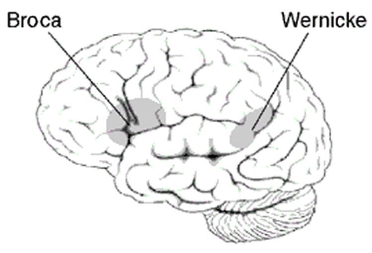
Based on language impairments caused by stroke, scientists believed a single area of the brain — a hotdog shaped section in the temporal lobe of the left hemisphere called Wernicke’s region — was the center of language comprehension. Wernicke’s was thought to be responsible for understanding the meaning of single words and sentences, two separate and critical functions.
But Northwestern Medicine scientists have updated and redrawn the traditional brain map of language comprehension based on new research with individuals who have a rare form of dementia that affects language, Primary Progressive Aphasia (PPA).
The new research shows word comprehension is actually located in a different brain neighborhood — the left anterior temporal lobe, a more forward location than Wernicke’s. And sentence comprehension turns out to be distributed widely throughout the language network, not in a single area as previously thought.
The paper was published June 25 in Brain.
“This provides an important change in our understanding of language comprehension in the brain,” said lead study author Dr. Marek-Marsel Mesulam, director of Northwestern’s Cognitive Neurology and Alzheimer’s Disease Center.
Mesulam also is a professor of neurology at Northwestern University Feinberg School of Medicine and a neurologist at Northwestern Memorial Hospital.
Knowing where language comprehension is located offers a more precise target for future therapies that could potentially protect or restore language function.
The stroke connection
Strokes cut off blood supply to regions of the brain and cause destruction of both neurons and fiber pathways passing through that region.
In the 1870s, a scientist named Carl Wernicke observed a specific region damaged by stroke and resulting language impairments. This area, consequently named Wernicke’s region, was identified as the seat of language comprehension.
“People who had strokes that affected Wernicke’s region couldn’t explain what a word such as umbrella meant,” Mesulam said. “Secondly, they had difficulty understanding sentence construction. If you said, ‘Put the apple on top of the book,’ even if they understood the meaning of apple and book, they wouldn’t be able to carry out the command because they can’t understand the construction of the sentence.”
Something doesn’t add up
But Mesulam, the world’s leading expert in PPA, for years had been puzzling over the fact that his PPA patients with damage in Wernicke’s area did not have the word comprehension impairment seen in stroke patients. They still understood individual words. And their sentence comprehension was inconsistent; some understood sentences; some didn’t.
“It was becoming clear over the many years I saw these patients, that there was some disconnect between what textbooks said and what we saw in our patients,” Mesulam said. “We did this study to analyze the discrepancy. The view of brain as seen from stroke did not match the view of the brain when seen from PPA.”
He and colleagues began a study of PPA patients, conducting quantitative MRI imaging of their brains and testing their language.
Northwestern scientist Emily Rogalski conducted the imaging in 72 PPA patients with damage inside and outside of Wernicke’s area. She measured cortex thickness in all of these areas. Cortex thickness is an indirect measure of the number of neurons and brain health. Thinning of the cortex in PPA indicates the destruction of neurons by the disease.
PPA patients still understand words
Rogalski, a research associate professor, found PPA patients who lost cortical thickness in Wernicke’s area still could understand individual words and had varied impairment of sentence comprehension. None of these patients had the global type of comprehension impairment described in stroke patients with Wernicke’s aphasia.
Severe word comprehension loss was only seen in PPA patients who had diminished cortical thickness in a region of the brain completely outside of Wernicke’s area, in the front part of the temporal lobe. This part of the brain is not prone to the effects of stroke, so its role in comprehension had been missed in prior language maps.
The discrepancy between the traditional map of comprehension and what was seen in PPA can be explained by the different ways the two diseases injure Wernicke’s area. In PPA, the neurodegenerative disease does not destroy the underlying fiber pathways that allow language areas to work together. But, in stroke patients, those critical highways passing through Wernicke’s had been blown up. So, the messages from other parts of the brain to the left anterior temporal lobe — the spot for word comprehension — were simply not getting through, Mesulam posits.
“What is happening here is no different from the charting of galaxies in outer space,” Mesulam said. “You look through one kind of telescope, you see one picture; you look through another infrared telescope, you get another picture. We are all in this pursuit of how to piece together different perspectives to get a better sense of how the brain works.”
“In this case, we saw a different map of language by comparing two different models of disease, one based on strokes that destroy an entire region of brain, cortex as well as underlying pathways, and the other on a neurodegenerative disease that attacks mostly brain cells in cortex rather than the region as a whole,” Mesulam said.
Other Northwestern authors on the paper are Sandra Weintraub and Cynthia Thompson.
Funding: The research was supported by grants DC008552 and DC01948 from the National Institute on Deafness and Communication Disorders, AG13854 from the National Institute on Aging, and NS075075 from the National Institute on Neurological Disease and Stroke, all of the National Institutes of Health. –
Source: Marla Paul – Northwestern University
Image Credit: Image credited to the NIH and is in the public domain
Original Research: Abstract for “The Wernicke conundrum and the anatomy of language comprehension in primary progressive aphasia” by M-Marsel Mesulam, Cynthia K. Thompson, Sandra Weintraub, and Emily J. Rogalski in Brain. Published online June 25 2015 doi:10.1093/brain/awv154
Abstract
The Wernicke conundrum and the anatomy of language comprehension in primary progressive aphasia
Wernicke’s aphasia is characterized by severe word and sentence comprehension impairments. The location of the underlying lesion site, known as Wernicke’s area, remains controversial. Questions related to this controversy were addressed in 72 patients with primary progressive aphasia who collectively displayed a wide spectrum of cortical atrophy sites and language impairment patterns. Clinico-anatomical correlations were explored at the individual and group levels. These analyses showed that neuronal loss in temporoparietal areas, traditionally included within Wernicke’s area, leave single word comprehension intact and cause inconsistent impairments of sentence comprehension. The most severe sentence comprehension impairments were associated with a heterogeneous set of cortical atrophy sites variably encompassing temporoparietal components of Wernicke’s area, Broca’s area, and dorsal premotor cortex. Severe comprehension impairments for single words, on the other hand, were invariably associated with peak atrophy sites in the left temporal pole and adjacent anterior temporal cortex, a pattern of atrophy that left sentence comprehension intact. These results show that the neural substrates of word and sentence comprehension are dissociable and that a circumscribed cortical area equally critical for word and sentence comprehension is unlikely to exist anywhere in the cerebral cortex. Reports of combined word and sentence comprehension impairments in Wernicke’s aphasia come almost exclusively from patients with cerebrovascular accidents where brain damage extends into subcortical white matter. The syndrome of Wernicke’s aphasia is thus likely to reflect damage not only to the cerebral cortex but also to underlying axonal pathways, leading to strategic cortico-cortical disconnections within the language network. The results of this investigation further reinforce the conclusion that the left anterior temporal lobe, a region ignored by classic aphasiology, needs to be inserted into the language network with a critical role in the multisynaptic hierarchy underlying word comprehension and object naming.
“The Wernicke conundrum and the anatomy of language comprehension in primary progressive aphasia” by M-Marsel Mesulam, Cynthia K. Thompson, Sandra Weintraub, and Emily J. Rogalski in Brain. Published online June 25 2015 doi:10.1093/brain/awv154