Since the mid-1800s, doctors have used drugs to induce general anesthesia in patients undergoing surgery. Despite their widespread use, little is known about how these drugs create such a profound loss of consciousness.
In a new study that tracked brain activity in human volunteers over a two-hour period as they lost and regained consciousness, researchers from MIT and Massachusetts General Hospital (MGH) have identified distinctive brain patterns associated with different stages of general anesthesia. The findings shed light on how one commonly used anesthesia drug exerts its effects, and could help doctors better monitor patients during surgery and prevent rare cases of patients waking up during operations.
Anesthesiologists now rely on a monitoring system that takes electroencephalogram (EEG) information and combines it into a single number between zero and 100. However, that index actually obscures the information that would be most useful, according to the authors of the new study, which appears in the Proceedings of the National Academy of Sciences.

“When anesthesiologists are taking care of someone in the operating room, they can use the information in this article to make sure that someone is unconscious, and they can have a specific idea of when the person may be regaining consciousness,” says senior author Emery Brown, an MIT professor of brain and cognitive sciences and health sciences and technology and an anesthesiologist at MGH.
Lead author of the paper is Patrick Purdon, an instructor of anesthesia at MGH and Harvard Medical School.
Distinctive patterns
Last fall, Purdon, Brown and colleagues published a study of brain activity in epileptic patients as they went under anesthesia. Using electrodes that had been implanted in the patients’ brains as part of their treatment for epilepsy, the researchers were able to identify a signature EEG pattern that emerged during anesthesia.
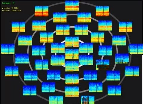
In the new study, the researchers studied healthy volunteers, measuring their brain activity with an array of 64 electrodes attached to the scalp. Not only did they find patterns that appeared to correspond to what they saw in last year’s study, they were also able to discern much more detail, because they gave the dose of propofol over a longer period of time and followed subjects until they came out of anesthesia.
While the subjects received propofol, the researchers monitored their responsiveness to sounds. Every four seconds, the subjects heard either a mechanical tone or a word, such as their name. The researchers measured EEG activity throughout the process, as the subjects pressed a button to indicate whether they heard the sound.
As the subjects became less responsive, distinct brain patterns appeared. Early on, when the subjects were just beginning to lose consciousness, the researchers detected an oscillation of brain activity in the low frequency (0.1 to 1 hertz) and alpha frequency (8 to 12 hertz) bands, in the frontal cortex. They also found a specific relationship between the oscillations in those two frequency bands: Alpha oscillations peaked as the low-frequency waves were at their lowest point.
When the brain reached a slightly deeper level of anesthesia, a marked transition occurred: The alpha oscillations flipped so their highest points occurred when the low frequency waves were also peaking.
The researchers believe that these alpha and low-frequency oscillations, which they also detected in last year’s study, produce unconsciousness by disrupting normal communication between different brain regions. The oscillations appear to constrain the amount of information that can pass between the frontal cortex and the thalamus, which normally communicate with each other across a very broad frequency band to relay sensory information and control attention.
The oscillations also prevent different parts of the cortex from coordinating with each other. In last year’s study, the researchers found that during anesthesia, neurons within small, localized brain regions are active for a few hundred milliseconds, then shut off again for a few hundred milliseconds. This flickering of activity, which creates the slow oscillation pattern, prevents brain regions from communicating normally.
Better anesthesia monitoring
When the researchers began to slowly decrease the dose of propofol, to bring the subjects out of anesthesia, they saw a reversal of the brain activity patterns that appeared when the subjects lost consciousness. A few minutes before regaining consciousness, the alpha oscillations flipped so that they were at their peak when the low-frequency waves were at their lowest point.
“That is the signature that would allow someone to determine if a patient is coming out of anesthesia too early, with this drug,” Purdon says.
Cases in which patients regain consciousness during surgery are alarming but very rare, with one or two occurrences in 10,000 operations, Brown says.
“It’s not something that we’re fighting with every day, but when it does happen, it creates this visceral fear, understandably, in the public. And anesthesiologists don’t have a way of responding because we really don’t know when you’re unconscious,” he says. “This is now a solved problem.”
Purdon and Brown are now starting a training program for anesthesiologists and residents at MGH to train them to interpret the information necessary to measure depth of anesthesia. That information is available through the EEG monitors that are now used during most operations, Purdon says. Because propofol is the most widely used anesthesia drug, the new findings should prove valuable for most operations.
Michael Ramsay, the chief of anesthesiology at Baylor University Medical Center, says he believes the new findings will improve patient care. The current monitoring system “has never been that accurate, and no one’s ever been able to show that it prevents awareness,” says Ramsay, who was not part of the research team. “What they have done is really brought back the value of interpreting the EEG signal that you’re looking at.”
In follow-up studies, the researchers are now studying the brain activity patterns produced by other anesthesia drugs.
Notes about this neurology research
The research was funded by the National Institutes of Health, including an NIH Director’s Pioneer Award, New Innovator Award and K-Award, and the Harvard Clinical and Translational Science Center.
Contact: Anne Trafton – MIT
Source: MIT press release
Image Source: The image is adapted from the video and is credited to Aylin Cimenser at MIT.
Video Source: The video, “MIT visualizes how the brain loses and regains consciousness” is available on the MIT News Video YouTube page.
Original Research: Full open access research (PDF and HTML) for “Electroencephalogram signatures of loss and recovery of consciousness from propofol” by Patrick L. Purdon, Eric T. Pierce, Eran A. Mukamel, Michael J. Prerau, John L. Walsh, Kin Foon K. Wong, Andres F. Salazar-Gomez, Priscilla G. Harrell, Aaron L. Sampson, Aylin Cimenser, ShiNung Ching, Nancy J. Kopell, Casie Tavares-Stoeckel, Kathleen Habeeb, Rebecca Merhar and Emery N. Brown in Proceedings of the National Academy of Sciences. Published online March 4 2013 doi:10.1073/pnas.1221180110