

Summary: Researchers discover smaller hippocampal gray matter volume in people who expressed higher or lower than average levels of cortisol in response to stress.
Source: SfN.
Individual differences in the pattern of release of the hormone cortisol in response to a stressful experience reveal how stressed a person actually feels, suggests a study of healthy women published in The Journal of Neuroscience. This approach could help to better identify and treat individuals more susceptible to the negative feelings associated with the physiological stress response.
Most stress research in humans involves inducing a short period of stress and classifying participants as either responders or non-responders based on the level of the hormone cortisol in their saliva. These studies do not typically find differences between the two groups’ subjective experience of stress.
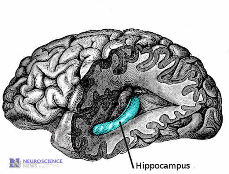
Roee Admon, Diego Pizzagalli and colleagues modified existing laboratory procedures to induce stress for more than one hour in 79 women and identified three different types of cortisol response. Participants that released either very high or very low amounts of cortisol over time reported feeling more stressed than those with a pattern of moderate hormonal release. The authors also found smaller volume of the hippocampus — a structure with a large number of cortisol receptors that regulates stress response — in these high and low responders compared to the moderate responders.
Source: David Barnstone – SfN
Image Source: NeuroscienceNews.com image is in the public domain.
Original Research: Abstract for “Distinct trajectories of cortisol response to prolonged acute stress are linked to affective responses and hippocampal gray matter volume in healthy females” by Roee Admon, Michael T. Treadway, Linda Valeri, Malavika Mehta, Samuel Douglas and Diego A. Pizzagalli in Journal of Neuroscience. Published online July 24 2017 doi:10.1523/JNEUROSCI.1175-17.2017
[cbtabs][cbtab title=”MLA”]SfN “Stress Hormone Cortisol Linked to Hippocampal Volume and Mood.” NeuroscienceNews. NeuroscienceNews, 24 July 2017.
<https://neurosciencenews.com/hippocampal-volume-cortisol-7159/>.[/cbtab][cbtab title=”APA”]SfN (2017, July 24). Stress Hormone Cortisol Linked to Hippocampal Volume and Mood. NeuroscienceNew. Retrieved July 24, 2017 from https://neurosciencenews.com/hippocampal-volume-cortisol-7159/[/cbtab][cbtab title=”Chicago”]SfN “Stress Hormone Cortisol Linked to Hippocampal Volume and Mood.” https://neurosciencenews.com/hippocampal-volume-cortisol-7159/ (accessed July 24, 2017).[/cbtab][/cbtabs]
Abstract
Distinct trajectories of cortisol response to prolonged acute stress are linked to affective responses and hippocampal gray matter volume in healthy females
The development of robust laboratory procedures for acute stress induction over the last decades has greatly advanced our understanding of stress responses in humans and their underlying neurobiological mechanisms. Nevertheless, attempts to uncover linear relationships between endocrine, neural, and affective responses to stress have generally yielded inconsistent results. Here, 79 healthy females completed a well-established laboratory procedure of acute stress induction that was modified in order to prolong its effect. Endocrinological and subjective affect assessments revealed stress-induced increases in cortisol release and negative affect that persisted 65 and 100 minutes post stress onset, respectively, confirming a relatively prolonged acute stress induction. Applying latent class linear mixed modelling (LCMM) on individuals’ patterns of cortisol responses identified three distinct trajectories of cortisol response: hyper-response (n=10), moderate-response (n=21), and mild-response (n=48). Notably, while all three groups exhibited a significant stress-induced increase in cortisol release and negative affect, hyper-response and mild-response groups both reported more negative affect relative to the moderate-response group. Structural MRI revealed no group differences in hippocampal and amygdala volumes, yet a continuous measure of cortisol response (area under the curve) showed that high and low levels of stress-induced cortisol release were associated with less hippocampal gray matter volume compared to moderate cortisol release. Together, these results suggest that distinct trajectories of cortisol response to prolonged acute stress among healthy females may not be captured by conventional linear analyses; instead, quadratic relations may better describe links between cortisol response to stress and affective responses, as well as hippocampal structural variability.
SIGNIFICANCE STATEMENT
Despite substantial research, it is unclear if and how individual neuroendocrine stress response patterns are linked to affective responses to stress and structural variability in neuroendocrine regulatory brain regions. By applying latent class linear mixed modelling on individuals’ patterns of cortisol responses to a prolonged acute stressor, we identified three distinct trajectories of cortisol response. Relative to the group showing moderate cortisol response, groups characterized by hyper and mild cortisol response were both associated with more negative affect. Moreover, a continuous measure of cortisol response showed that high and low levels of stress-induced cortisol release correlated with reduced hippocampal gray matter volume. Given that neuroendocrine stress responses are conceptualized as biomarkers of stress susceptibility, these insights may have clinical implications.
“Distinct trajectories of cortisol response to prolonged acute stress are linked to affective responses and hippocampal gray matter volume in healthy females” by Roee Admon, Michael T. Treadway, Linda Valeri, Malavika Mehta, Samuel Douglas and Diego A. Pizzagalli in Journal of Neuroscience. Published online July 24 2017 doi:10.1523/JNEUROSCI.1175-17.2017


