Dogs have a specialized region in their brains for processing faces, a new study finds. PeerJ is publishing the research, which provides the first evidence for a face-selective region in the temporal cortex of dogs.
“Our findings show that dogs have an innate way to process faces in their brains, a quality that has previously only been well-documented in humans and other primates,” says Gregory Berns, a neuroscientist at Emory University and the senior author of the study.
Having neural machinery dedicated to face processing suggests that this ability is hard-wired through cognitive evolution, Berns says, and may help explain dogs’ extreme sensitivity to human social cues.
Berns heads up the Dog Project in Emory’s Department of Psychology, which is researching evolutionary questions surrounding man’s best, and oldest, friend. The project was the first to train dogs to voluntarily enter a functional magnetic resonance imaging (fMRI) scanner and remain motionless during scanning, without restraint or sedation.
In previous research, the Dog Project identified the caudate region of the canine brain as a reward center. It also showed how that region of a dog’s brain responds more strongly to the scents of familiar humans than to the scents of other humans, or even to those of familiar dogs.
For the current study, the researchers focused on how dogs respond to faces versus everyday objects. “Dogs are obviously highly social animals,” Berns says, “so it makes sense that they would respond to faces. We wanted to know whether that response is learned or innate.”
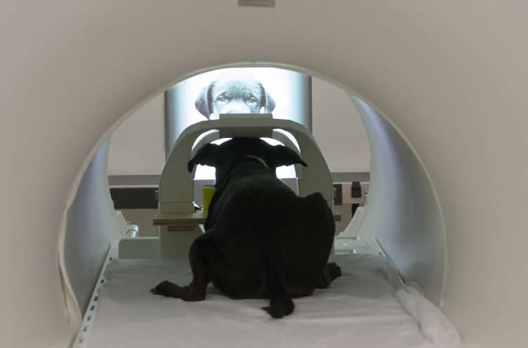
The study involved dogs viewing both static images and video images on a screen while undergoing fMRI. It was a particularly challenging experiment since dogs do not normally interact with two-dimensional images, and they had to undergo training to learn to pay attention to the screen.
A limitation of the study was the small sample size: Only six of the eight dogs enrolled in the study were able to hold a gaze for at least 30 seconds on each of the images to meet the experimental criteria.
The results were clear, however, for the six subjects able to complete the experiment. A region in their temporal lobe responded significantly more to movies of human faces than to movies of everyday objects. This same region responded similarly to still images of human faces and dog faces, yet significantly more to both human and dog faces than to images of everyday objects.
If the dogs’ response to faces was learned – by associating a human face with food, for example – you would expect to see a response in the reward system of their brains, but that was not the case, Berns says.
A previous study, decades ago, using electrophysiology, found several face-selective neurons in sheep.
“That study identified only a few face-selective cells and not an entire region of the cortex,” says Daniel Dilks, an Emory assistant professor of psychology and the first author of the current dog study.
The researchers have dubbed the canine face-processing region they identified the dog face area, or DFA.
Humans have at least three face processing regions in the brain, including the fusiform face area, or FFA, which is associated with distinguishing faces from other objects. “We can predict what parts of your brain are going to be activated when you’re looking at faces,” Dilks says. “This is incredibly reliable across people.”
One hypothesis is that distinguishing faces is important for any social creature.
“Dogs have been cohabitating with humans for longer than any other animal,” Dilks says. “They are incredibly social, not just with other members of their pack, but across species. Understanding more about canine cognition and perception may tell us more about social cognition and perception in general.”
Source: Carol Clark – Emory University
Image Credit: The image is credited to Gregory Berns, Emory University
Original Research: Full open access research for “Awake fMRI reveals a specialized region in dog temporal cortex for face processing” by Daniel D. Dilks, Peter Cook, Samuel K. Weiller, Helen P. Berns, Mark Spivak, and Gregory S. Berns in PeerJ. Published online August 4 2015 doi:10.7717/peerj.1115
Abstract
Awake fMRI reveals a specialized region in dog temporal cortex for face processing
Recent behavioral evidence suggests that dogs, like humans and monkeys, are capable of visual face recognition. But do dogs also exhibit specialized cortical face regions similar to humans and monkeys? Using functional magnetic resonance imaging (fMRI) in six dogs trained to remain motionless during scanning without restraint or sedation, we found a region in the canine temporal lobe that responded significantly more to movies of human faces than to movies of everyday objects. Next, using a new stimulus set to investigate face selectivity in this predefined candidate dog face area, we found that this region responded similarly to images of human faces and dog faces, yet significantly more to both human and dog faces than to images of objects. Such face selectivity was not found in dog primary visual cortex. Taken together, these findings: (1) provide the first evidence for a face-selective region in the temporal cortex of dogs, which cannot be explained by simple low-level visual feature extraction; (2) reveal that neural machinery dedicated to face processing is not unique to primates; and (3) may help explain dogs’ exquisite sensitivity to human social cues.
“Awake fMRI reveals a specialized region in dog temporal cortex for face processing” by Daniel D. Dilks, Peter Cook, Samuel K. Weiller, Helen P. Berns, Mark Spivak, and Gregory S. Berns in PeerJ. Published online August 4 2015 doi:10.7717/peerj.1115