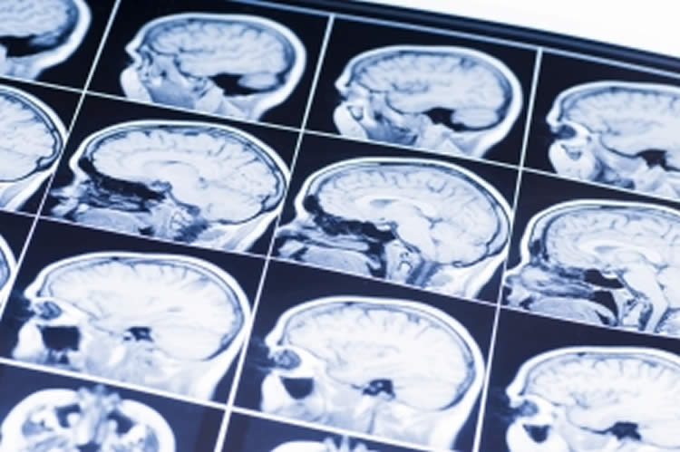
Summary: Researchers report an MRI test to measure smaller volumes in specific hippocampal regions could be used to detect Alzheimer’s disease.
Source: McGill University.
New research has drawn a link between changes in the brain’s anatomy and biomarkers that are known to appear at the earliest stages of Alzheimer’s disease (AD), findings that could one day provide a sensitive but non-invasive test for AD before cognitive symptoms appear.
Scientists have known for some time that one of the first signs of AD is buildup of amyloid-Beta and tau proteins in the brain. They have also known that the hippocampus atrophies and loses volume in some AD patients years before cognitive decline.
To examine the link between the two, a team of researchers from McGill University and McGill-affiliated health institutes followed 88 individuals at hereditary risk of AD, but who did not show any cognitive signs of the disease. Subjects were scanned using magnetic resonance imaging (MRI) to determine brain volume and had cerebrospinal fluid (CSF) extracted to test levels of amyloid-Beta and tau. These data were collected by the Centre for Studies on the Prevention of Alzheimer’s Disease (StoP-AD) group at the Douglas Mental Health University Institute under the leadership of Dr. John Breitner.
Using statistical models, the researchers found that the high levels of both tau and amyloid-Beta are associated with smaller volumes and image intensity profiles of specific regions of the hippocampal circuit, and that this is less likely when there is accumulation of one protein but not the other. The intensity related finding suggests that researchers can use MRI to examine changes occurring at a microstructural level that may even precede more severe volume deficits.
Biomarkers can be used to test the effectiveness of drugs in clinical trials
Their results add to our knowledge of how AD progresses from its first physiological signs to cognitive decline, helping identify those at most risk of AD. These biomarkers can then be used to test the effectiveness of drugs in clinical trials. It also may one day allow physicians to identify at-risk people with only MRI, eliminating the need for a lumbar puncture, which can be a painful procedure.
“Our work highlights not only the need but also the possibility of adding sensitive biomarkers of early white matter pathology in the presymptomatic phase of AD,” says Christine Tardif, an assistant professor at the McConnell Brain Imaging Centre of the Montreal Neurological Institute and Hospital, and the paper’s first author.
“This technique demonstrates significant promise in identifying those at greatest risk for developing Alzheimer’s disease without using an invasive procedure like a lumbar puncture, which can be stressful for patients” says Mallar Chakravarty, the study’s senior author and an assistant professor at McGill’s Department of Psychiatry and computational neuroscientist in the Cerebral Imaging Centre at the Douglas Mental Health University Institute.
Funding: This study was funded with generous support from the StoP-AD Centre.
Source: Shawn Hayward – McGill University
Publisher: Organized by NeuroscienceNews.com.
Image Source: NeuroscienceNews.com image is adapted from the McGill University news release.
Original Research: Abstract for “Regionally specific changes in the hippocampal circuitry accompany progression of cerebrospinal fluid biomarkers in preclinical Alzheimer’s disease” by Tardif CL, Devenyi GA, Amaral RSC, Pelleieux S, Poirier J, Rosa-Neto P, Breitner J, Chakravarty MM; and PREVENT-AD Research Group in Human Brain Mapping. Published online November 21 2017 doi:10.1002/hbm.23897
[cbtabs][cbtab title=”MLA”]McGill University “A Non-Invasive Method to Detect Alzheimer’s.” NeuroscienceNews. NeuroscienceNews, 20 December 2017.
<https://neurosciencenews.com/alzheimers-brain-volume-8217/>.[/cbtab][cbtab title=”APA”]McGill University (2017, December 20). A Non-Invasive Method to Detect Alzheimer’s. NeuroscienceNews. Retrieved December 20, 2017 from https://neurosciencenews.com/alzheimers-brain-volume-8217/[/cbtab][cbtab title=”Chicago”]McGill University “A Non-Invasive Method to Detect Alzheimer’s.” https://neurosciencenews.com/alzheimers-brain-volume-8217/ (accessed December 20, 2017).[/cbtab][/cbtabs]
Abstract
Regionally specific changes in the hippocampal circuitry accompany progression of cerebrospinal fluid biomarkers in preclinical Alzheimer’s disease
Neuropathological and in vivo brain imaging studies agree that the cornu ammonis 1 and subiculum subfields of the hippocampus are most vulnerable to atrophy in the prodromal phases of Alzheimer’s disease (AD). However, there has been limited investigation of the structural integrity of the components of the hippocampal circuit, including subfields and extra-hippocampal white matter structure, in relation to the progression of well-accepted cerebrospinal fluid (CSF) biomarkers of AD, amyloid-β 1-42 (Aβ) and total-tau (tau). We investigated these relationships in 88 aging asymptomatic individuals with a parental or multiple-sibling familial history of AD. Apolipoprotein (APOE) ɛ4 risk allele carriers were identified, and all participants underwent cognitive testing, structural magnetic resonance imaging, and lumbar puncture for CSF assays of tau, phosphorylated-tau (p-tau) and Aβ. Individuals with a reduction in CSF Aβ levels (an indicator of amyloid accretion into neuritic plaques) as well as evident tau pathology (believed to be linked to neurodegeneration) exhibited lower subiculum volume, lower fornix microstructural integrity, and a trend towards lower cognitive score than individuals who showed only reduction in CSF Aβ. In contrast, persons with normal levels of tau showed an increase in structural MR markers in relation to declining levels of CSF Aβ. These results suggest that hippocampal subfield volume and extra-hippocampal white matter microstructure demonstrate a complex pattern where an initial volume increase is followed by decline among asymptomatic individuals who, in some instances, may be a decade or more away from onset of cognitive or functional impairment.
“Regionally specific changes in the hippocampal circuitry accompany progression of cerebrospinal fluid biomarkers in preclinical Alzheimer’s disease” by Tardif CL, Devenyi GA, Amaral RSC, Pelleieux S, Poirier J, Rosa-Neto P, Breitner J, Chakravarty MM; and PREVENT-AD Research Group in Human Brain Mapping. Published online November 21 2017 doi:10.1002/hbm.23897