

Neuronal feedback could change what we ‘see’.
Ever see something that isn’t really there? Could your mind be playing tricks on you? The “tricks” might be your brain reacting to feedback between neurons in different parts of the visual system, according to a study published in the Journal of Neuroscience by Carnegie Mellon University Assistant Professor of Biological Sciences Sandra J. Kuhlman and colleagues.
Understanding this feedback system could provide new insight into the visual system’s neuronal circuitry and could have further implications for understanding how the brain interprets and understands sensory stimuli.
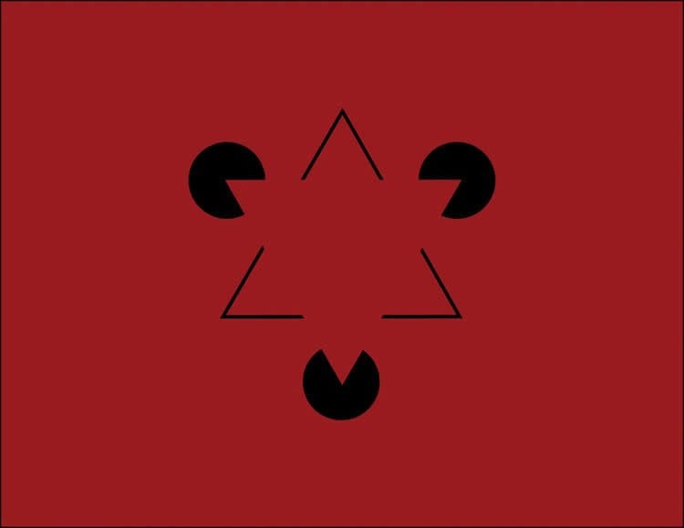
Many optical illusions make you see something that’s not there. Take the Kanizsa triangle: when you place three Pac-Man-like wedges in the right spot, you see a triangle, even though the edges of the triangle aren’t drawn.
“We see with both our brain and our eyes. Your brain is making inferences that allow you to see the triangle. It’s connecting the dots between the corners of the wedges,” said Kuhlman, who is a member of Carnegie Mellon’s BrainHub neuroscience initiative and the joint Carnegie Mellon/University of Pittsburgh Center for the Neural Basis of Cognition (CNBC). “Optical illusions illustrate some of the amazing things our visual system can do.”
When we look at an object, information about what we see travels through circuits of neurons beginning in the retina, through the thalamus and into the brain’s visual cortex. In the visual cortex, the information gets processed in multiple stages and is ultimately sent to the prefrontal cortex — the area of the brain that makes decisions, including how to respond to a given stimulus.
However, not all information stays on this forward moving path. At the secondary stage of processing in the visual cortex some neurons reverse course and send information back to the first stage of processing. Researchers at Carnegie Mellon wondered if this feedback could change how the neurons in the visual cortex respond to a stimulus and alter the messages being sent to the prefrontal cortex.
While there has been a good deal of research studying how information moves forward through the visual system, less has been done to study the impact of the information that moves backward. To find out if the information traveling from the secondary stage of processing back to the first stage impacted how information is encoded in the visual system, the researchers needed to quantify the magnitude of information that was being sent from the second stage back to the first stage. Using a mouse model, they recorded normal neuronal firing in the first stage of the visual cortex as the mouse looked at moving patterns that represented edges. They then silenced the neurons in the second stage using modified optogenetic technology. This halted the feedback of information from the second stage back to the first stage, and allowed the researchers to determine how much of the neuronal activity in the first stage of visual processing was the result of feedback.
Twenty percent of the neuronal activity in the visual cortex was the result of feedback, a concept Kuhlman calls reciprocal connectivity. This indicates that some of the information coming from the visual cortex is not a direct response to a visual stimuli, but is a response to how the stimuli was perceived by higher cortical areas.
The feedback, she says, might be what causes our brain to complete the undrawn lines in the Kanizsa triangle. But more importantly, it signifies that studying neuronal feedback is important to our understanding of how the brain works to process stimuli.
“This represents a new way to study visual perception and neural computation. If we want to truly understand the visual pathway, and cortical function in general, we have to understand these reciprocal connection,” Kuhlman said.
Additional authors on this study are Carnegie Mellon and the CNBC’s Diego E. Pafundo, Mark A. Nicholas and Ruilin Zhang.
Funding: This study was funded by the Knights Templar Eye Foundation, the Howard Hughes Medical Institute Undergraduate Program, the Fight-For-Sight Foundation and the National Institutes of Health’s National Eye Institute (R01-EY024678).
Source: Jocelyn Duffy – Carnegie Mellon University
Image Source: The image adapted from the Carnegie Mellon University press release.
Original Research: Abstract for “Top-Down-Mediated Facilitation in the Visual Cortex Is Gated by Subcortical Neuromodulation” by Diego E. Pafundo, Mark A. Nicholas, Ruilin Zhang, and Sandra J. Kuhlman in Journal of Neuroscience. Published online March 9 2016 doi:10.1523/JNEUROSCI.2909-15.2016
Abstract
Top-Down-Mediated Facilitation in the Visual Cortex Is Gated by Subcortical Neuromodulation
Response properties in primary sensory cortices are highly dependent on behavioral state. For example, the nucleus basalis of the forebrain plays a critical role in enhancing response properties of excitatory neurons in primary visual cortex (V1) during active exploration and learning. Given the strong reciprocal connections between hierarchically arranged cortical regions, how are increases in sensory response gain constrained to prevent runaway excitation? To explore this, we used in vivo two-photon guided cell-attached recording in conjunction with spatially restricted optogenetic photo-inhibition of higher-order visual cortex in mice. We found that the principle feedback projection to V1 originating from the lateral medial area (LM) facilitated visual responses in layer 2/3 excitatory neurons by ∼20%. This facilitation was reduced by half during basal forebrain activation due to differential response properties between LM and V1. Our results demonstrate that basal-forebrain-mediated increases in response gain are localized to V1 and are not propagated to LM and establish that subcortical modulation of visual cortex is regionally distinct.
SIGNIFICANCE STATEMENT Reciprocal connectivity among brain regions is a prominent feature of all sensory cortices. In primary visual cortex (V1), top-down signals from association areas aid in context-dependent perception of visual scenes by altering the response properties of individual neurons. Sensory-evoked responses in V1 are also highly dependent on subcortical neuromodulation pathways that regulate brain state. Here, with cell-type-specific resolution, we addressed how corticocortical and subcortical pathways interact to regulate responsiveness of V1. Our results provide insight into the rules and conditions governing activity propagation in reciprocally connected networks.
“Top-Down-Mediated Facilitation in the Visual Cortex Is Gated by Subcortical Neuromodulation” by Diego E. Pafundo, Mark A. Nicholas, Ruilin Zhang, and Sandra J. Kuhlman in Journal of Neuroscience. Published online March 9 2016 doi:10.1523/JNEUROSCI.2909-15.2016


