Summary: Researchers made a breakthrough in understanding memory loss due to repeated head impacts, as often experienced by athletes. Their study reveals that memory issues following head injury in mice are linked to inadequate reactivation of neurons involved in memory formation.
This discovery is significant because it demonstrates that the memory loss is not a permanent, degenerative condition but potentially reversible. By using lasers to activate specific memory neurons, the researchers successfully reversed amnesia in mice, opening new avenues for treating cognitive impairments in humans caused by repeated head impacts.
Key Facts:
- The study shows that memory loss from head impacts is related to the failure to reactivate specific memory-forming neurons, not permanent damage.
- Researchers successfully reversed memory loss in mice using laser activation of memory neurons.
- This research provides a new understanding of memory issues in athletes and others with repeated head impacts, suggesting potential for non-invasive human treatments.
Source: Georgetown University
A mouse study designed to shed light on memory loss in people who experience repeated head impacts, such as athletes, suggests the condition could potentially be reversed. The research in mice finds that amnesia and poor memory following head injury is due to inadequate reactivation of neurons involved in forming memories.
The study, conducted by researchers at Georgetown University Medical Center in collaboration with Trinity College Dublin, Ireland, is reported January 16, 2024, in the Journal of Neuroscience.
Importantly for diagnostic and treatment purposes, the researchers found that the memory loss attributed to head injury was not a permanent pathological event driven by a neurodegenerative disease. Indeed, the researchers could reverse the amnesia to allow the mice to recall the lost memory, potentially allowing cognitive impairment caused by head impact to be clinically reversed.
The Georgetown investigators had previously found that the brain adapts to repeated head impacts by changing the way the synapses in the brain operate. This can cause trouble in forming new memories and remembering existing memories. In their new study, investigators were able to trigger mice to remember memories that had been forgotten due to head impacts.
“Our research gives us hope that we can design treatments to return the head-impact brain to its normal condition and recover cognitive function in humans that have poor memory caused by repeated head impacts,” says the study’s senior investigator, Mark Burns, PhD, a professor and Vice-Chair in Georgetown’s Department of Neuroscience and director of the Laboratory for Brain Injury and Dementia.
In the new study, the scientists gave two groups of mice a new memory by training them in a test they had never seen before. One group was exposed to a high frequency of mild head impacts for one week (similar to contact sport exposure in people) and one group were controls that didn’t receive the impacts. The impacted mice were unable to recall the new memory a week later.
“Most research in this area has been in human brains with chronic traumatic encephalopathy (CTE), which is a degenerative brain disease found in people with a history of repetitive head impact,” said Burns. “By contrast, our goal was to understand how the brain changes in response to the low-level head impacts that many young football players regularly experience.”
Researchers have found that, on average, college football players receive 21 head impacts per week with defensive ends receiving 41 head impacts per week. The number of head impacts to mice in this study were designed to mimic a week of exposure for a college football player, and each single head impact by itself was extraordinarily mild.
Using genetically modified mice allowed the researchers to see the neurons involved in learning new memories, and they found that these memory neurons (the “memory engram”) were equally present in both the control mice and the experimental mice.
To understand the physiology underlying these memory changes, the study’s first author, Daniel P. Chapman, Ph.D., said, “We are good at associating memories with places, and that’s because being in a place, or seeing a photo of a place, causes a reactivation of our memory engrams. This is why we examined the engram neurons to look for the specific signature of an activated neuron.
“When the mice see the room where they first learned the memory, the control mice are able to activate their memory engram, but the head impact mice were not. This is what was causing the amnesia.”
The researchers were able to reverse the amnesia to allow the mice to remember the lost memory using lasers to activate the engram cells.
“We used an invasive technique to reverse memory loss in our mice, and unfortunately this is not translatable to humans,” Burns adds.
“We are currently studying a number of non-invasive techniques to try to communicate to the brain that it is no longer in danger, and to open a window of plasticity that can reset the brain to its former state.”
In addition to Burns and Chapman the authors include Stefano Vicini at Georgetown University and Sarah D. Power and Tomás J. Ryan at Trinity College Dublin, Ireland.
Funding: This work was supported by the Mouse Behavior Core in the Georgetown University Neuroscience Department and by the National Institutes of Health (NIH) / National Institute of Neurological Disorders and Stroke (NINDS) grants R01NS107370 & R01NS121316. NINDS also supported F30 NS122281 and the Neural Injury and Plasticity Training Grant housed in the Center for Neural Injury and Recovery at Georgetown University (T32NS041218). Seed funding is from the CTE Research Fund at Georgetown.
The authors report having no personal financial interests related to the study.
About this TBI and neurology research news
Author: Karen Teber
Source: Georgetown University
Contact: Karen Teber – Georgetown University
Image: The image is credited to Neuroscience News
Original Research: Closed access.
“Amnesia after repeated head impact is caused by impaired synaptic plasticity in the memory engram” by Mark Burns et al. Journal of Neuroscience
Abstract
Amnesia after repeated head impact is caused by impaired synaptic plasticity in the memory engram
Sub-concussive head impacts are associated with the development of acute and chronic cognitive deficits. We recently reported that high-frequency head impact (HFHI) causes chronic cognitive deficits in mice through synaptic changes. To better understand the mechanisms underlying HFHI-induced memory decline, we used TRAP2/Ai32 transgenic mice to enable visualization and manipulation of memory engrams.
We labeled the fear memory engram in male and female mice exposed to an aversive experience and subjected them to sham or HFHI. Upon subsequent exposure to natural memory recall cues, sham, but not HFHI mice, successfully retrieved fearful memories.
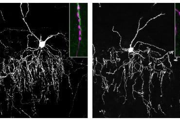
In sham mice the hippocampal engram neurons exhibited synaptic plasticity, evident in amplified AMPA:NMDA ratio, enhanced AMPA-weighted tau, and increased dendritic spine volume compared to non-engram neurons. In contrast, although HFHI mice retained a comparable number of hippocampal engram neurons, these neurons did not undergo synaptic plasticity.
This lack of plasticity coincided with impaired activation of the engram network, leading to retrograde amnesia in HFHI mice. We validated that the memory deficits induced by HFHI stem from synaptic plasticity impairments by artificially activating the engram using optogenetics, and found that stimulated memory recall was identical in both sham and HFHI mice.
Our work shows that chronic cognitive impairment after HFHI is a result of deficiencies in synaptic plasticity instead of a loss in neuronal infrastructure, and we can reinstate a forgotten memory in the amnestic brain by stimulating the memory engram. Targeting synaptic plasticity may have therapeutic potential for treating memory impairments caused by repeated head impacts.


