Summary: For decades, scientists have known that the protein tau acts like a wildfire in the brain, leaping from region to region and leaving a trail of cognitive decline. Now, a decade-long study has finally mapped the mechanism of this spread.
By combining fMRI brain scans of living patients with postmortem analysis of their brain tissue, researchers discovered that tau “seeds” travel primarily along an individual’s unique, natural communication pathways. This confirms that personal neuronal wiring dictates how fast and far Alzheimer’s advances, providing a definitive target for antibody therapies designed to “catch” tau as it moves between synapses.
Key Facts
- The Seed Theory: Tau begins as “seeds” that travel between neurons through synapses (the connection points where brain cells “talk”). Once a seed reaches a new neuron, it triggers the formation of neurofibrillary tangles.
- The Study Scale: Researchers analyzed data from 128 participants over 10 years, integrating genetic data, antemortem fMRI (scans taken while alive), and postmortem “seed bioactivity” (analysis after death).
- Individual Specificity: Because every person’s brain is wired differently, the path tau takes is unique to each individual. Your “connectome” (brain map) determines your specific risk and progression speed.
- Proving Causality: Using a statistical method called Mendelian Randomization, the team proved that tau seeds generated in the temporal cortex (memory center) directly cause the destructive tangles found later in the neocortex (complex thought center).
- Validation for Antibodies: These findings explain why tau-targeting antibodies show promise in clinical trials; by intercepting tau as it travels outside the cell between synapses, these drugs can effectively “break the chain” of infection.
Source: UAB
Alzheimer’s disease is a progressive neurodegenerative disorder that slowly impairs memory, affects thinking skills, and eventually interferes with daily functioning. It is the most common cause of dementia, which affects millions of families around the world and places a vast emotional and economic toll on families.
In Alzheimer’s disease, two key proteins, extracellular amyloid-beta plaques and an intercellular protein called tau, disrupt communication between the brain’s cells and lead to cell damage and death.
As tau becomes abnormal it forms neurofibrillary tangles and spreads through critical regions of the brain, triggering cell death and the cognitive decline characteristic of Alzheimer’s disease.
A study published in Neuron led by researchers at the University of Alabama at Birmingham, Rush University Medical Center in Chicago, Illinois, and SUNY Upstate Medical Center in Syracuse, New York, provides new insight into a fundamental mystery of Alzheimer’s disease: how tau tangles spread from one brain region to another.
The findings of the study introduce evidence that targeting tau as it spreads can offer a viable path to slowing or preventing the advancement of Alzheimer’s disease.
Tau is a microtubule-associated protein that is found inside the brain’s neurons that helps support their internal structure by serving as a “scaffolding.” In people with Alzheimer’s disease, however, tau proteins begin sticking together inside cells.
These tangles clump together and impair neural function, which eventually kills the cells. The more that tau spreads, the more memory loss that occurs. What remained unclear was the mechanism by which tau travels through the brain’s network.
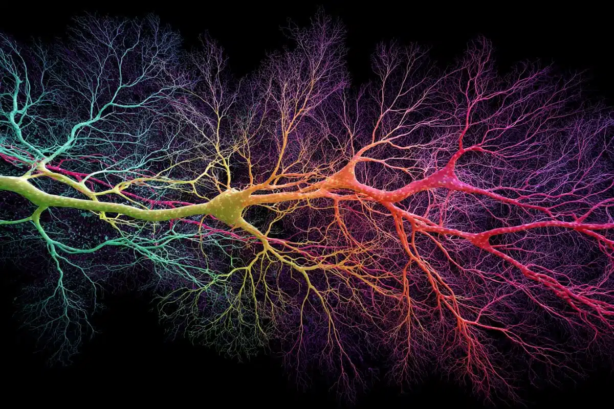
“Small pieces of tau make up the aggregate inside the neuron and spread from neuron to neuron through the brain. Neurons are connected to each other, and they talk to each other through synapses.
“This would allow them to get around the brain and deposit and aggregate at different brain sections until it reaches the neocortex, “said Jeremy Herskowitz, Ph.D., professor of neurology and neurobiology at UAB, and the Patsy W. and Charles A. Collat professor of neuroscience, and corresponding author of this study.
“While that’s a theory; our paper shows that’s the likely the mechanism of action as people age.”
“This is a major advancement in Alzheimer’s research for both therapy development and understanding how the disease works,” said Herskowitz.
Herskowitz and his team analyzed postmortem brain samples and longitudinal data from 128 ROSMAP participants, a Rush University study involving Catholic clergy ages 65+ who undergo annual evaluations and donate their brains after death. The participants averaged 91 years old at death, with nearly one‑third having Alzheimer’s dementia. Gathering the fMRI and postmortem data for this research took a decade.
Researchers examined two brain samples from each participant. One sample was from the lower temporal lobe, which is vital for memory recall. The second was from the upper frontal lobe, which supports working memory and complex thought. Tau typically begins accumulating in the temporal lobe before spreading to the frontal lobe. This progression mirrors the shift from early memory problems to more advanced cognitive decline.
The researchers integrated multiple types of information from the ROSMAP participants. First, they looked at tau seeds in both brain regions alongside each person’s genetic data to determine if tau seeds caused the formation of tau tangles. They used a method called Mendelian randomization, which helps determine cause and effect.
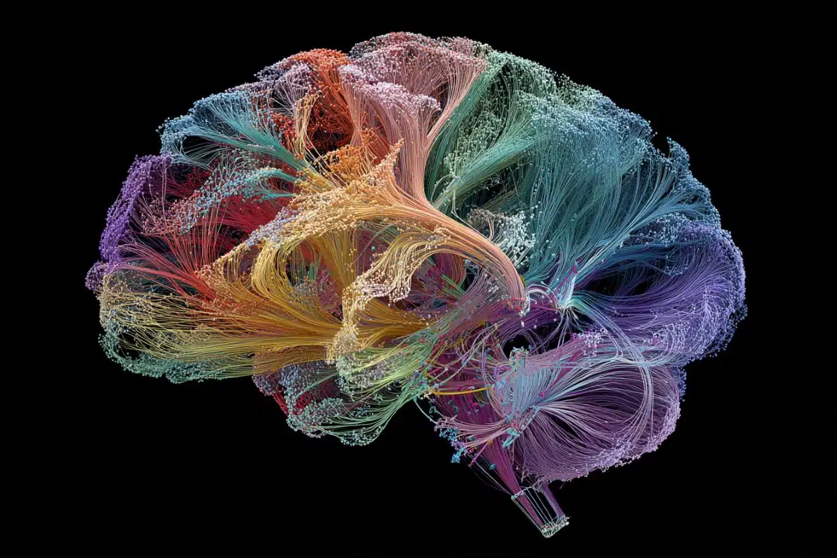
Brain-connectivity data called antemortem functional magnetic resonance imaging (fMRI) was used to examine each individual’s unique pattern of brain wiring and evaluate whether personal wiring differences influence the amount of tau seed tangles and how far the tangles spread.
“We used a genetic approach called Mendelian Causality to make the conclusion that the seeds that were generated in the temporal cortex caused the neurofibrillary tangle pathology in the neocortex. The Mendelian causality that we used is a statistical algorithm that utilizes the genomic DNA that we had from each participant, and that allowed us to make this conclusion,” said Herskowitz.
This study represents the largest investigation of tau seed bioactivity in human brains to date, which has never been paired with fMRI data. Overall results indicate significant findings. The researchers discovered that tau seeds could spread primarily along an individual’s natural communication pathways.
These pathways, neurons, connect at synapses which form a vast network that varies from person to person. Tau seeds appear to travel along these pathways, moving from one synapse to the next and seeding new tangles as they go. In short, an individual’s unique brain connectivity helps determine how far and how fast tau pathology advances.
While future research is necessary to fully explore the specific mechanisms used by tau seeds to spread across the cerebral cortex through synapses, the study clearly demonstrates that personal differences in neuronal wiring influence the spread of Alzheimer’s pathology. Collectively, these findings reinforce the therapeutic potential of targeting tau seeds to slow or halt Alzheimer’s disease progression.
Previous clinical trials have shown that tau traveling outside of the cell has been susceptible to being targeted by therapeutic antibodies. With these novel research findings, the reason for the antibody’s success is now known.
“Tau antibodies would stop tau from spreading from one brain region to the next. If you stop that spreading, it would delay or prevent Alzheimer’s disease dementia,” said Herskowitz.
First author of the study “Tau seeds induce neurofibrillary tangle formation across brain regions via individual specific connectivity” is Audrey Weber, Ph.D., UAB Department of Neurology. At UAB, neurology and neurobiology are departments in the Heersink School of Medicine.
Other authors of the study are Kelsey M. Greathouse, UAB Department of Neurology and Center for Neurodegeneration and Experimental Therapies; David A. Bennett and Shinya Tasaki, Rush University Medical Center; and Chris Gaiteri, and Bernard Ng, Rush University Medical Center and SUNY Upstate Medical University.
Funding: Support came from National Institutes of Health grants F99AG083305, T32NS095775, R21AG085379, P30AG086401, R01AG061800, R01AG061798, R01AG057911, U01AG079847, P30AG10161, P30AG72975, R01AG15819, R01AG17917, U01AG46152, U01AG61356.
Key Questions Answered:
A: In a way, yes. While you can’t “catch” it from someone else, the protein tau behaves like a slow-moving infection inside the head. It “seeds” itself in one healthy neuron, hitches a ride across the synapse to the next, and forces the healthy proteins there to misfold and clump together.
A: Most studies only look at the brain after the damage is done. By using fMRI data from when the participants were alive, researchers could see the “roads” (neural pathways) before they were destroyed. This allowed them to prove that tau followed those specific roads to reach other parts of the brain.
A: That is the goal. Since the study shows tau follows your personal “wiring,” future doctors might be able to use a brain scan to predict exactly which regions are at risk next and intervene with personalized antibody treatments to block those specific “highways.”
Editorial Notes:
- This article was edited by a Neuroscience News editor.
- Journal paper reviewed in full.
- Additional context added by our staff.
About this Alzheimer’s disease research news
Author: Rachel Beatty
Source: UAB
Contact: Rachel Beattya – UAB
Image: The image is credited to Neuroscience News
Original Research: Open access.
“Tau seeds induce neurofibrillary tangle formation across brain regions via individual-specific connectivity” by Audrey J. Weber, Bernard Ng, Kelsey M. Greathouse, David A. Bennett, Shinya Tasaki, Chris Gaiteri, and Jeremy H. Herskowitz. Neuron
DOI:10.1016/j.neuron.2026.03.001
Abstract
Tau seeds induce neurofibrillary tangle formation across brain regions via individual-specific connectivity
The spread of tau pathology across the cerebral cortex is closely tied to cognitive decline in Alzheimer’s disease (AD).
To investigate mechanisms underlying tau spread, we measured bioactivity of tau seeds from inferior temporal gyrus (ITG) and superior frontal gyrus (SFG) synaptosomes in 128 individuals and demonstrated that tau seed bioactivity associates with tau phosphorylation, neurofibrillary tangles (NFTs), and cognitive impairment.
Incorporating genotype data from the same individuals within a Mendelian randomization framework showed that tau seeds in ITG induce NFTs locally as well as drive tau seeds and NFTs in SFG.
Integrating antemortem functional magnetic resonance imaging data from the same individuals showed that person-specific connectivity modulates the tau seed-NFT relationships.
These findings indicate that tau seeds underlie the spread of NFTs both locally and across distal brain regions via individual-specific connectivity.