

Summary: A specialized area of the mouse brain called the SuM specializes in detecting novel experiences. Within this brain area, responses to social novelty, or experiences related to unknown individuals, were segregated from those related to unfamiliar places, before being sent to areas of the brain associated with memory.
Source: RIKEN
In a study published in the scientific journal Nature, researchers at the RIKEN Center for Brain Science (CBS) in Japan report that a part of the mouse brain called the SuM is specialized for detecting new experiences. Within the SuM, responses to experiences related to unknown individuals–called social novelty–were segregated from those related to unfamiliar places–called context novelty–before being sent to distinct parts of the brain’s main memory-formation center.
This discovery can help us understand normal memory, as well as conditions in which recognizing and reacting to new information is impaired.
Meeting someone for the first time or entering an unfamiliar apartment is a much different experience than meeting an acquaintance or walking into your own home. Normal social interactions, daily functions, and even survival can depend on being able to make the distinction between the unknown and the familiar. Almost all animals seem to be born with this ability, and like other innate behaviors, the team at RIKEN CBS hypothesized that a region of the brain called the hypothalamus might be involved.
To test this hypothesis, they exposed mice to two types of novelty: contextual or social. The novel context was an unfamiliar cage with a few landmark objects and the social novelty was an unfamiliar juvenile mouse. They found that overall brain activity in a part of the hypothalamus called the SuM was much higher in these novel situations than when mice were placed in familiar cages or near familiar mice.
“The hypothalamus is a very highly conserved region of the brain across evolution, mostly thought to be involved in innate behaviors like feeding, mating, parenting, and fighting,” says Team Leader Thomas McHugh.
“Our data suggest that it could also serve as a link between these survival-type behaviors and higher cognitive function.”
Surprisingly, although the SuM signaled novelty overall, the majority of individual brain cells in the SuM only responded to one or the other type of novel situation. This is the first time that anyone has found a social/contextual split within a novelty circuit in the brain.
In order to see how far the separation went, the team needed to create a new transgenic mouse line that would allow them to see exactly where these SuM neurons project and what they do.
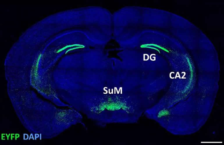
A series of experiments showed that the neurons in the SuM connect with two parts of the hippocampus, the part of the brain known for being involved in memory formation and storage. Neurons that were selective for contextual novelty connected to the DG part of the hippocampus, while those that signaled social novelty were connected to the CA2 region.
Scientists often use novelty tests to assess memory. Mice explore new places and approach unfamiliar mice, making these behaviors both signs of novelty, and by inference, a lack of memory.
McHugh and his team used optogenetic light stimulation to show that output from the SuM directly affected social and contextual memory.
For example, exciting the SuM-to-CA2 connection with blue light caused mice to behave as if they had a selective deficit in social memory; they frequently approached familiar mice as if they had never seen them before, but did not explore familiar rooms more than control mice.
Likewise, they saw the reverse behavioral pattern when the SuM-to-DG connection was excited.
Accurately detecting contextual and social novelty allows us to adapt our behavior appropriately to changes in daily life.
“Understanding how we recognize and react to novel information is fundamental to understanding memory,” says McHugh.
“Not only does novelty strengthen memory, both in mice and humans, impairment in recognizing and reacting to new information often accompanies psychiatric conditions. This research can thus provide a biological target to examine in such cases.”
About this genetics research article
Source:
Garvan Institute of Medical Research
Contacts:
Adam Phillips – RIKEN
Image Source:
The image is credited to RIKEN.
Original Research: Closed access
“A hypothalamic novelty signal modulates hippocampal memory” by Thomas J. McHugh et al. Nature.
Abstract
A hypothalamic novelty signal modulates hippocampal memory
The ability to recognize information that is incongruous with previous experience is critical for survival. Novelty signals have therefore evolved in the mammalian brain to enhance attention, perception and memory. Although the importance of regions such as the ventral tegmental area and locus coeruleus in broadly signalling novelty is well-established, these diffuse monoaminergic transmitters have yet to be shown to convey specific information on the type of stimuli that drive them. Whether distinct types of novelty, such as contextual and social novelty, are differently processed and routed in the brain is unknown. Here we identify the supramammillary nucleus (SuM) as a novelty hub in the hypothalamus. The SuM region is unique in that it not only responds broadly to novel stimuli, but also segregates and selectively routes different types of information to discrete cortical targets—the dentate gyrus and CA2 fields of the hippocampus—for the modulation of mnemonic processing. Using a new transgenic mouse line, SuM-Cre, we found that SuM neurons that project to the dentate gyrus are activated by contextual novelty, whereas the SuM–CA2 circuit is preferentially activated by novel social encounters. Circuit-based manipulation showed that divergent novelty channelling in these projections modifies hippocampal contextual or social memory. This content-specific routing of novelty signals represents a previously unknown mechanism that enables the hypothalamus to flexibly modulate select components of cognition.


