Summary: Study reveals brain wave activity in the medial prefrontal cortex and amygdala associated with social behavior in mice.
Source: Tohoku University
Researchers at Tohoku University and the University of Tokyo have discovered electrical wave patterns in the brain related to social behavior in mice. They also observed that mice showing signs of stress, depression, or autism lacked these brain waves.
The medial prefrontal cortex (mPFC) and amygdala regions of the brain regulate our emotion, and undergo pathological changes when we experience psychiatric diseases. However, the detailed neuronal processes behind this remain unclear.
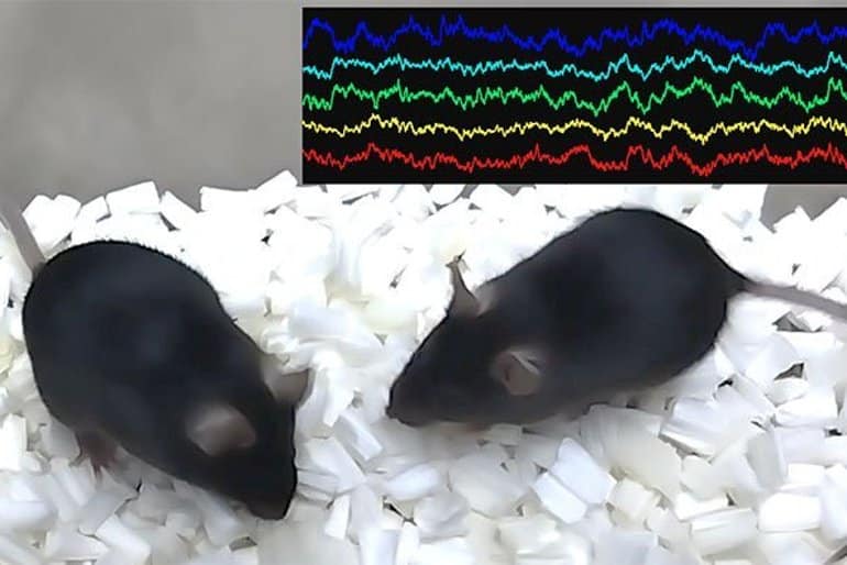
Takuya Sasaki from Tohoku University’s Graduate School of Pharmaceutical Sciences led a collaborative team who recorded electrical brain signals—so-called brain electrical waves—in the mPFC and amygdala areas of mice.

They found that certain brain waves underwent pronounced variations when the mice interacted socially with one another. Specifically, brain waves at the frequency band of theta (4-7 Hz) and gamma (30-60 Hz) decreased and increased, respectively, during socializing.
When the same tests were applied to mice exhibiting poor social skills or symptoms of depression and autism, the brain waves were not present. Notably, artificially replicating social behavior-related brain waves by an optical and genetic manipulation technique in these pathological mouse models restored their ability to interact socially.
“This finding provides a unified understanding of brain activity underlying social behavior and its deficits in disease,” says Sasaki.
Looking ahead, Sasaki is eager to identify the basic mechanisms of neuronal dynamics in these brain waves and evaluate the involvement of the other brain regions in social behavior. In conjunction, he is investigating whether the same brain mechanisms work in humans for clinical applications.
About this social behavior research news
Author: Press Office
Source: Tohoku University
Contact: Press Office – Tohoku University
Image: The image is credited to Takuya Sasaki et al.
Original Research: Open access.
“Prefrontal-amygdalar oscillations related to social behavior in mice” by Nahoko Kuga et al. eLife
Abstract
Prefrontal-amygdalar oscillations related to social behavior in mice
The medial prefrontal cortex and amygdala are involved in the regulation of social behavior and associated with psychiatric diseases but their detailed neurophysiological mechanisms at a network level remain unclear.
We recorded local field potentials (LFPs) from the dorsal medial prefrontal cortex (dmPFC) and basolateral amygdala (BLA) while male mice engaged on social behavior. We found that in wild-type mice, both the dmPFC and BLA increased 4–7 Hz oscillation power and decreased 30–60 Hz power when they needed to attend to another target mouse.
In mouse models with reduced social interactions, dmPFC 4–7 Hz power further increased especially when they exhibited social avoidance behavior. In contrast, dmPFC and BLA decreased 4–7 Hz power when wild-type mice socially approached a target mouse. Frequency-specific optogenetic manipulations replicating social approach-related LFP patterns restored social interaction behavior in socially deficient mice.
These results demonstrate a neurophysiological substrate of the prefrontal cortex and amygdala related to social behavior and provide a unified pathophysiological understanding of neuronal population dynamics underlying social behavioral deficits.


