

Summary: According to a new study, the brains of people with schizophrenia may be able to reorganize and fight the illness.
Source: Lawson Health Research Institute.
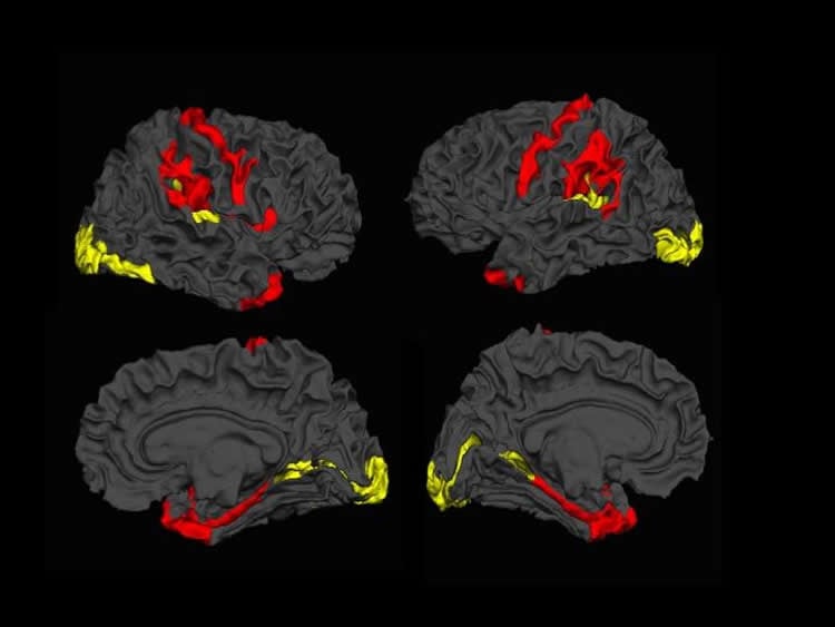
ncrease in the brain’s grey matter proof that the brain has the ability to rescue itself.
A team of scientists from across the globe have shown that the brains of patients with schizophrenia have the capacity to reorganize and fight the illness. This is the first time that imaging data has been used to show that our brains may have the ability to reverse the effects of schizophrenia.
Schizophrenia is an illness generally associated with a widespread reduction in brain tissue volume. However, a recent study found that a subtle increase in tissue also occurs in certain brain regions.
The study followed 98 patients with schizophrenia and compared them to 83 patients without schizophrenia. The team used Magnetic Resonance Imaging (MRI) and a sophisticated approach called covariance analysis to record the amount of brain tissue increase. Due to the subtlety and the distributed nature of increase, this had not been demonstrated in patients before now.
According to Lawson Health Research Institute’s Dr. Lena Palaniyappan, there is an overarching feeling that curing people with a severe mental illness, such as schizophrenia is not possible. “Even the state-of-art frontline treatments aim merely for a reduction rather than a reversal of the cognitive and functional deficits caused by the illness,” says Dr. Palaniyappan, who is the Medical Director at the Prevention & Early Intervention Program for Psychoses (PEPP) at London Health Sciences Centre (LHSC).
This comes from a long-standing notion that schizophrenia is a degenerative illness, with the seeds of damage sown very early during the course of brain development. “Our results highlight that despite the severity of tissue damage, the brain of a patient with schizophrenia is constantly attempting to reorganize itself, possibly to rescue itself or limit the damage,” says Dr. Palaniyappan.
The team’s next step is to clarify the evolution of this brain tissue reorganization process by repeatedly scanning individual patients with early schizophrenia and study the effect of this reorganization on their recovery.
“These findings are important not only because of their novelty and the rigour of the study, but because they point the way to the development of targeted treatments that potentially could better address some of the core pathology in schizophrenia,” explains Dr. Jeffrey Reiss, Site Chief, Psychiatry, LHSC. “Brain plasticity and the development of related therapies would contribute to a new optimism in an illness that was 100 years ago described as premature dementia for its seemingly progressive deterioration.”
“Dr. Palaniyappan and his colleagues have opened new avenues of research into our understanding of schizophrenia,” says Dr. Paul Links, Chair/Chief, Psychiatry, LHSC. “Their findings may lead us to be able to harness the brain’s own compensatory changes in the face of this illness and improve recovery. We are excited that Dr. Palaniyappan will be continuing this important clinical research here in London with his international colleagues.”
The project is the result of an international collaboration among scientists in Nottingham, UK, Shanghai and Changsha, People’s Republic of China, Robarts Research Institutes at Western University and Lawson Health Research Institute.
The study, “Dynamic cerebral reorganization in the pathophysiology of schizophrenia: a MRI-derived cortical thickness study” is published online in the current issue of Psychology Medicine.
Source: Julia Capaldi – Lawson Health Research Institute
Image Source: This NeuroscienceNews.com image is credited to Lena Palaniyappan.
Original Research: Abstract for “Dynamic cerebral reorganization in the pathophysiology of schizophrenia: a MRI-derived cortical thickness study” by S. Guo, L. Palaniyappan, P. F. Liddle and J. Feng in Psychological Medicine. Published online May 26 2016 doi:10.1017/S0033291716000994
[cbtabs][cbtab title=”MLA”]Lawson Health Research Institute. “Imaging Study Shows Promising Results for People With Schizophrenia.” NeuroscienceNews. NeuroscienceNews, 28 May 2016.
<https://neurosciencenews.com/schizophrenia-gray-matter-neuroimaging-4332/>.[/cbtab][cbtab title=”APA”]Lawson Health Research Institute. (2016, May 28). Imaging Study Shows Promising Results for People With Schizophrenia. NeuroscienceNews. Retrieved May 28, 2016 from https://neurosciencenews.com/schizophrenia-gray-matter-neuroimaging-4332/[/cbtab][cbtab title=”Chicago”]Lawson Health Research Institute. “Imaging Study Shows Promising Results for People With Schizophrenia.” https://neurosciencenews.com/schizophrenia-gray-matter-neuroimaging-4332/ (accessed May 28, 2016).[/cbtab][/cbtabs]
Abstract
Dynamic cerebral reorganization in the pathophysiology of schizophrenia: a MRI-derived cortical thickness study
Background A structural neuroanatomical change indicating a reduction in brain tissue is a notable feature of schizophrenia. Several pathophysiological processes such as aberrant cortical maturation, progressive tissue loss and compensatory tissue increase could contribute to the structural changes seen in schizophrenia.
Method We studied cortical thickness using surface-based morphometry in 98 clinically stable patients with schizophrenia and 83 controls. Using a pattern classification approach, we studied whether the features that discriminate patients from controls vary across the different stages of the illness. Using a covariance analysis, we also investigated if concurrent increases accompany decreases in cortical thickness.
Results Very high levels of accuracy (96.3%), specificity (98.8%) and sensitivity (88%) were noted when classifying patients with <2 years of illness from controls. Within the patient group, reduced thickness was consistently accompanied by increased thickness in distributed brain regions. A pattern of cortical amelioration or normalization (i.e. reduced deviation from controls) was noted with increasing illness duration. While temporo-limbic and fronto-parietal regions showed reduced thickness, the occipital cortex showed increased thickness, especially in those with a long-standing illness.
Conclusion A compensatory remodelling process might contribute to the cortical thickness variations in different stages of schizophrenia. Subtle cerebral reorganization reflecting the inherent plasticity of brain may occur concomitantly with processes contributing to tissue reduction in adult patients with schizophrenia.
“Dynamic cerebral reorganization in the pathophysiology of schizophrenia: a MRI-derived cortical thickness study” by S. Guo, L. Palaniyappan, P. F. Liddle and J. Feng in Psychological Medicine. Published online May 26 2016 doi:10.1017/S0033291716000994


