Summary: Blood can bring more oxygen into the brains of mice following exercise as increased respiration increases oxygen levels in hemoglobin.
Source: Penn State
Contrary to accepted knowledge, blood can bring more oxygen to mice brains when they exercise because the increased respiration packs more oxygen into the hemoglobin, according to an international team of researchers who believe that this holds true for all mammals.
“Standard thought was that mammalian blood is always completely saturated with oxygen,” said Patrick J. Drew, Huck Distinguished Associate Professor of Neural Engineering and Neurosurgery and associate director of the Penn State Neuroscience Institute.
That would mean that the only way to get more oxygen to the brain would be to get more blood to the brain by increasing blood flow. The researchers were interested in seeing how brain oxygen levels were affected by natural behaviors, specifically exercise.
“We know that people change breathing patterns when doing cognitive tasks,” said Drew. “In fact, respiration phase locks to the task at hand. In the brain, increases in neural activity usually are accompanied by increases in blood flow.”
However, exactly what is happening in the body was unknown, so the researchers used mice who could chose to walk or run on a treadmill and monitored their respiration, neural activity, blood flow and brain oxygenation.
“We predicted that brain oxygenation would depend on neural activity and blood flow,” said Qing Guang Zhang, postdoctoral fellow in engineering science and mechanics. “We expected the oxygenation would drop in the brain’s frontal cortex if blood flow decreased.
“That was what we thought would happen, but then we realized it was the respiration that was keeping the oxygenation up.”
The only way that could happen would be if exercise was causing the blood to carry more oxygen, he explained, which would mean that the blood was not normally completely saturated with oxygen.
The researchers looked at oxygenation in the somatosensory cortex and the frontal cortex — which is an area involved in cognition — and the olfactory bulb — an area involved in the sense of smell — because they are the most accessible areas of the brain.

They used a variety of methods to monitor respiration, blood flow and oxygenation. They also tested oxygenation levels while suppressing neural activity and blood vessel dilation.
The researchers report in today’s (Dec. 4) issue of Nature Communications that “The oxygenation persisted when neural activity and functional hyperemia (blood flow increases) were blocked, occurred both in the tissue and in arteries feeding the brain, and were tightly correlated with respiration rate and the phase of respiration cycle.”
They conclude that “respiration provides a dynamic pathway for modulating cerebral oxygenation.”
Also working on this project at Penn State were Kyle W. Gheres, graduate student in molecular, cellular and integrative biosciences; Ravi Kedrasetti, doctoral student in engineering science and mechanics; and William D. Haselden, M.D./Ph.D. student in the Medical Scientist Training Program and Neuroscience Graduate Program.
Others on the project include Morgane Roche, graduate student; Emmanuelle Chaigneau, postdoctoral researcher; and Serge Charpak, professor of neuroscience, all at the Institut de la Santé et de la Recherche Médicale, Paris, France.
Funding: The McKnight Endowment Fund for Neuroscience and the National Institutes of Health supported this research.
Source:
Penn State
Media Contacts:
A’ndrea Elyse Messer – Penn State
Image Source:
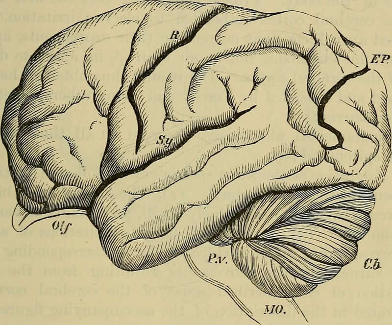
The image is credited to Handbook of Physiology, 1892 William Morrant Baker.
Original Research: Open access
“Cerebral oxygenation during locomotion is modulated by respiration”. Qingguang Zhang, Morgane Roche, Kyle W. Gheres, Emmanuelle Chaigneau, Ravi T. Kedarasetti, William D. Haselden, Serge Charpak & Patrick J. Drew.
Nature Communications doi:10.1038/s41467-019-13523-5.
Abstract
Cerebral oxygenation during locomotion is modulated by respiration
In the brain, increased neural activity is correlated with increases of cerebral blood flow and tissue oxygenation. However, how cerebral oxygen dynamics are controlled in the behaving animal remains unclear. We investigated to what extent cerebral oxygenation varies during locomotion. We measured oxygen levels in the cortex of awake, head-fixed mice during locomotion using polarography, spectroscopy, and two-photon phosphorescence lifetime measurements of oxygen sensors. We find that locomotion significantly and globally increases cerebral oxygenation, specifically in areas involved in locomotion, as well as in the frontal cortex and the olfactory bulb. The oxygenation increase persists when neural activity and functional hyperemia are blocked, occurred both in the tissue and in arteries feeding the brain, and is tightly correlated with respiration rate and the phase of respiration cycle. Thus, breathing rate is a key modulator of cerebral oxygenation and should be monitored during hemodynamic imaging, such as in BOLD fMRI.


