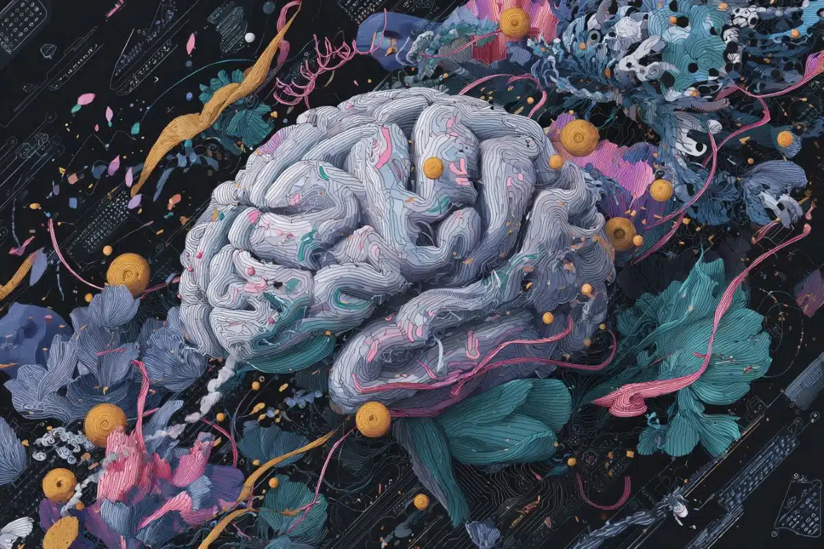
Summary: Protein synthesis in different amygdala neuron populations is essential for storing information about threat and safety.
Source: NYU
A team of neuroscientists has identified processes the brain undergoes to distinguish real and present dangers from those linked to past experiences in mice. The findings, which appear in the journal Nature, have implications for our understanding of post-traumatic stress disorder (PTSD)–an affliction marked by the inability to distinguish between past and present dangers or to recognize “safe” situations.
“Memories of a traumatic episode can last for a long time,” says Professor Eric Klann, director of New York University’s Center for Neural Science and the paper’s senior author. “But we are able to use such memories selectively: to predict and respond to a subsequent, related danger while also recognizing when threats do not exist. This is especially important for survival behavior in an uncertain environment such as a conflict zone or at times of social unrest.”
“This has significant implications for memory disorders such as PTSD, where patients have difficulty distinguishing between safety and threat cues,” adds lead author Prerana Shrestha, a postdoctoral researcher in NYU’s Center for Neural Science.
The study, which also included researchers from Rockefeller University and McGill University, focused on the neurological processes that mice use to make these distinctions.
Learning to identify and appropriately respond to cues in an uncertain environment is crucial for animal survival, the researchers note. Specifically, cues that reliably predict danger prompt behaviors such as freezing in order to escape detection. However, along with the threat-predicting cues, an uncertain environment can present cues that predict safety–or, specifically, lack of danger. Animals, then, need to respond to the threat-predicting cue with defensive behaviors and, conversely, to safety cues by ceasing a threat response and resuming normal behaviors.
In the Nature study, the scientists sought to identify the cellular molecules, or substrates, for long-term storage of threat and safety-cue-associated memories.
It has been long established that a region of the brain, the amygdala, plays a fundamental role in the processing and storing of emotion-related information. Less understood, however, are the cellular engines and architecture that underlie it–specifically, the identity of cell types that store cue-related information and allow animals to respond appropriately even after considerable time has elapsed after the initial threat exposure.
Also well understood are the formation and consolidation of long-lasting memories, which occur through changes in the cellular landscape of proteins–a dynamic that captures significant features of an event, in part by synthesis of new proteins.
In the new work, the scientists aimed to better understand these mechanisms by disrupting key steps in protein synthesis in specific cell types–a maneuver that would reveal their significance. This procedure allowed the researchers to identify key players in this intricate process.

To do so, they examined and perturbed the assembly of two protein complexes that are crucial for the synthesis of new proteins. The first protein complex contains eIF2, which is involved in adding the first amino acid to a protein being synthesized. The second protein complex contains eIF4E, which binds to the protected ‘cap’ of messenger RNA that is necessary for them to be translated into protein.
Notably, they found that protein synthesis in specific inhibitory neurons in the amygdala–Somatostatin-expressing neurons–is crucial for storing information about the cued threat whereas protein synthesis in PKCδ-expressing neurons is necessary for storing complementary information about safety cues.
Activity in these populations of neurons was previously shown to occur in processing threat-related cues; however, this is the first study to connect the necessity of new protein synthesis in these neurons to the stabilization of long-term emotional memories.
Funding: The research was funded by grants from the National Institutes of Health (R37-NS034007, R01-NS047384) and Brain and Behavior Research Foundation (26696).
In a related study also appearing in this issue of Nature, researchers at McGill University, the University of Montreal, and Haifa University looked at eIF2 in different types of neurons as well. They found that increasing the eIF2 protein complex in Somatostatin-expressing inhibitory neurons, which results in increased protein synthesis, boosts the consolidation of long-term memory.
Together, both studies illuminate previously unknown ways the eIF2 protein complex calibrates the strength of fearful memories.
About this neurotech research news
Source: AAAS
Contact: Press Office – AAAS
Image: The image is credited to B. Conlon et al., Science Translational Medicine (2020).
Original Research: Closed access.
“N-Acetylglucosamine drives myelination by triggering oligodendrocyte precursor cell differentiation” by Michael Demetriou et al. Journal of Biological Chemistry.
Abstract
Amygdala inhibitory neurons as loci for translation in emotional memories
To survive in a dynamic environment, animals need to identify and appropriately respond to stimuli that signal danger1. Survival also depends on suppressing the threat-response during a stimulus that predicts the absence of threat (safety). An understanding of the biological substrates of emotional memories during a task in which animals learn to flexibly execute defensive responses to a threat-predictive cue and a safety cue is critical for developing treatments for memory disorders such as post-traumatic stress disorder. The centrolateral amygdala is an important node in the neuronal circuit that mediates defensive responses, and a key brain area for processing and storing threat memories. Here we applied intersectional chemogenetic strategies to inhibitory neurons in the centrolateral amygdala of mice to block cell-type-specific translation programs that are sensitive to depletion of eukaryotic initiation factor 4E (eIF4E) and phosphorylation of eukaryotic initiation factor 2α (p-eIF2α). We show that de novo translation in somatostatin-expressing inhibitory neurons in the centrolateral amygdala is necessary for the long-term storage of conditioned-threat responses, whereas de novo translation in protein kinase Cδ-expressing inhibitory neurons in the centrolateral amygdala is necessary for the inhibition of a conditioned response to a safety cue. Our results provide insight into the role of de novo protein synthesis in distinct inhibitory neuron populations in the centrolateral amygdala during the consolidation of long-term memories.