Summary: White matter tracts were significantly altered in babies whose mothers experienced stress during pregnancy.
Source: King’s College London
New research from King’s College London has found that maternal stress before and during pregnancy could affect a baby’s brain development.
In their study published in Biological Psychiatry, MRC Doctoral Researcher in Perinatal Imaging and Health, Alexandra Lautarescu and Head of Advanced Neuroimaging, Professor Serena Counsell, for the first time looked at the relationship between maternal stress and brain development in 251 premature babies.
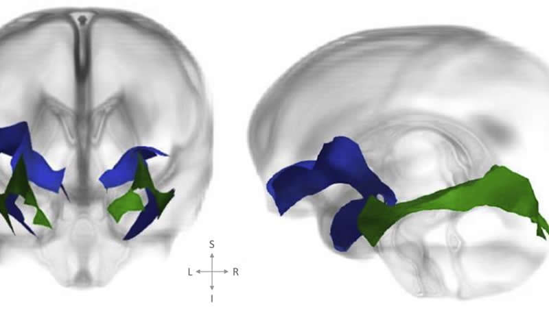
They found evidence for impaired development of a white matter tract, the uncinate fasciculus, in babies whose mothers experienced more stress in the prenatal period.
The mothers completed a questionnaire which asked them about their experiences of stressful events, which ranged from everyday stress such as moving house or taking an exam to more severe stressors like experiencing bereavement, separation or divorce. A score of severity of stress was calculated based on how many stressors they experienced as well as how severe those stressors were. This is what was related to the baby’s brain. The researchers used a medical imaging technique called diffusion tensor imaging that was specifically developed to look at the structure of the white matter. The white matter tract has previously been implicated in anxiety disorders – adults that have an anxiety disorder may show changes in this tract.
“We found that in the mums that were more stressed during pregnancy and the period before birth, white matter was altered in the babies,” said lead researcher Alexandra Lautarescu from King’s College London.
Scientists say the study highlights the importance of providing support for expectant mothers, as previous studies have shown that interventions such as cognitive behavioural therapy can help mitigate adverse outcomes in the baby. Clinicians have an important role to play when speaking with expectant mothers. While questions are asked about depressive symptoms, few questions are asked about general stress and anxiety. Women who deal with stressful life events during pregnancy are not picked up by their GPs or by their health care providers very often.
“It is not diagnosed as often as it should be during pregnancy and we are trying to emphasise that maternal mental health during pregnancy can impact the baby’s brain development which may impact on their outcomes later in life,” Alexandra Lautarescu said. “No one is asking these women about stress and hence they don’t receive any support.
“Antenatal services need to be aware that it is important to think about stress of the mums and we need to have some kind of support there for the mums who identify that they are stressed. If we try to help these women either during the pregnancy or in the early post-natal period with some sort of intervention this will not only help the mother, but may also prevent impaired brain development in the baby and improve their outcomes overall.”
There is some evidence to suggest that if mothers experience poor mental health during pregnancy that leads to adverse outcomes in the baby – obstetric outcomes, lower birth weight or premature birth. A mother’s poor mental health may also lead to altered early behavior such as more frequent crying.
Further studies are needed to understand whether the observed changes in the brain development of these babies will lead to adverse outcomes later in life.
Source:
King’s College London
Media Contacts:
Tanya Wood – King’s College London
Image Source:
The image is credited to Alexandra Lautarescu et al.
Original Research: Open access
“Maternal Prenatal Stress Is Associated With Altered Uncinate Fasciculus Microstructure in Premature Neonates”. Alexandra Lautarescu, Diliana Pecheva, Chiara Nosarti, Julie Nihouarn, Hui Zhang, Suresh Victor, Michael Craig, A. David Edwards, and others.
Biological Psychiatry doi:10.1016/j.biopsych.2019.08.010.
Abstract
Maternal Prenatal Stress Is Associated With Altered Uncinate Fasciculus Microstructure in Premature Neonates
Background
Maternal prenatal stress exposure (PNSE) increases risk for adverse psychiatric and behavioral outcomes in offspring. The biological basis for this elevated risk is poorly understood but may involve alterations to the neurodevelopmental trajectory of white matter tracts within the limbic system, particularly the uncinate fasciculus. Additionally, preterm birth is associated with both impaired white matter development and adverse developmental outcomes. In this study we hypothesized that higher maternal PNSE was associated with altered uncinate fasciculus microstructure in offspring.
Methods
In this study, 251 preterm infants (132 male, 119 female) (median gestational age = 30.29 weeks [range, 23.57–32.86 weeks]) underwent brain magnetic resonance imaging including diffusion-weighted imaging around term-equivalent age (median = 42.43 weeks [range, 37.86–45.71 weeks]). Measures of white matter microstructure were calculated for the uncinate fasciculus and the inferior longitudinal fasciculus, a control tract that we hypothesized was not associated with maternal PNSE. Multiple regressions were used to investigate the relationship among maternal trait anxiety scores, stressful life events, and white matter microstructure indices in the neonatal brain.
Results
Adjusting for gestational age at birth, postmenstrual age at scan, maternal age, socioeconomic status, sex, and number of days on parenteral nutrition, higher stressful life events scores were associated with higher axial diffusivity (β = .177, q = .007), radial diffusivity (β = .133, q = .026), and mean diffusivity (β = .149, q = .012) in the left uncinate fasciculus, and higher axial diffusivity (β = .142, q = .026) in the right uncinate fasciculus.
Conclusions
These findings suggest that PNSE is associated with altered development of specific frontolimbic pathways in preterm neonates as early as term-equivalent age.