Summary: Researchers have discovered how the brain keeps time for precise movements, revealing a neural “hourglass” mechanism between the motor cortex and striatum. The motor cortex sends timing signals that accumulate in the striatum until they reach a threshold that triggers action.
Temporarily disrupting either region pauses or resets this internal timer, directly altering movement timing. The findings offer major insight into how the brain coordinates actions and could inform therapies for motor disorders like Parkinson’s and Huntington’s disease.
Key Facts
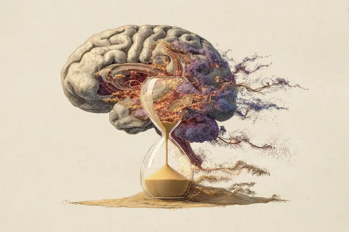
- Neural Hourglass: Timing signals flow from the motor cortex to the striatum, accumulating like sand in an hourglass.
- Pause vs. Reset: Silencing the motor cortex pauses the timer, while silencing the striatum resets it entirely.
- Therapeutic Potential: Understanding this timing system may lead to strategies for restoring movement control in motor disorders.
Source: Max Planck Institute
Whether speaking or swinging a bat, precise and adaptable timing of movement is essential for everyday behavior.
Although we do not have sensory organs like eyes or a nose to sense time, we can keep time and control the timing of our actions. Such timing accuracy depends on a timer in the brain, but how the brain implements this timer was previously unknown.
In research published this week in Nature, MPFI scientists Zidan Yang, Hidehiko Inagaki, and colleagues reveal how this timer works through the interaction of two brain regions—the motor cortex and the striatum.
Together, these areas track the passage of time much like an hourglass.
Discovering the Brain’s Hourglass
Prior studies on how the brain might time movement have highlighted both the motor cortex and the striatum as key brain regions. These regions show neural activity patterns consistent with timing functions and cause movement timing deficits when damaged in diseases such as Parkinson’s and Huntington’s.
Dr. Zidan Yang, lead scientist of the study, explains, “We understood there was an adjustable timer in the brain, but it was unclear how the brain was implementing this timer and what the specific role of each brain region was. We wanted to understand precisely how the brain keeps time because it is such a critical function for our everyday activities.”
To achieve this, the scientists trained mice to receive a treat by licking a spout with specific timing, for example, after 1 second. During this task, the researchers recorded the activity of thousands of neurons in both the motor cortex and the striatum to measure their timing-related patterns.
To understand how the brain’s timer might work, scientists combined these measurements with a technique called optogenetics, which allowed them to temporarily silence the activity of one brain area with flashes of light and measure the resulting changes in the timing-related patterns in the other area.
“By combining neural recordings with brief changes in the activity of specific brain regions, we were able to identify the role that each region played in the brain’s internal timer. We realized that these brain regions work together to track time, but play unique roles – similar to the top and bottom of an hourglass,” described Dr. Yang.
Pausing and Rewinding the Timer
The researchers discovered that the motor cortex is like the top of the hourglass, sending streams of neural signals to the striatum. In the striatum, those signals accumulate as time passes, just like the sand at the bottom of the hourglass. Once the signal reaches a certain level, movement is triggered.
When the researchers temporarily silenced the motor cortex, it paused the flow of these signals as if pinching the neck of the hourglass to stop the flow of sand. This paused the buildup of activity in the striatum and delayed the timing of the mouse’s lick for the treat, as if time itself had been paused.
On the other hand, when the researchers silenced the striatum, it reset the timing signals as if the hourglass had been flipped to restart the timer. This delayed the mouse’s licking even further, as if time had been rewound.
Broader Impacts
The team’s findings mark a significant advancement in understanding how neural activity across these two areas interact to coordinate the timing of actions. Dr. Hidehiko Inagaki, MPFI research group leader and senior author of the study, describes his ultimate goal:
“The motor cortex and striatum are the two key brain areas that control our movement and are damaged in many motor disorders. We are working to understand how the brain’s activity patterns across these critical brain areas lead to precise control of behavior, such as fluid movements. We hope that this understanding can be harnessed to restore movement functions to those facing the challenges of living with a motor disorder.”
Key Questions Answered:
A: By using coordinated activity between the motor cortex and striatum that accumulates signals like an hourglass.
A: Silencing the motor cortex pauses the timer, while silencing the striatum resets it entirely.
A: Because these regions are impaired in motor disorders, understanding this timer could guide new treatments.
Editorial Notes:
- This article was edited by a Neuroscience News editor.
- Journal paper reviewed in full.
- Additional context added by our staff.
About this movement and neuroscience research news
Author: Lesley Colgan
Source: Max Planck Institute
Contact: Lesley Colgan – Max Planck Institute
Image: The image is credited to Neuroscience News
Original Research: Open access.
“Integrator dynamics in the cortico-basal ganglia loop for flexible motor timing” by Zidan Yang et al. Nature
Abstract
Integrator dynamics in the cortico-basal ganglia loop for flexible motor timing
Flexible control of motor timing is crucial for behaviour. Before volitional movement begins, the frontal cortex and striatum exhibit ramping spiking activity, with variable ramp slopes anticipating movement onsets. This activity in the cortico-basal ganglia loop may function as an adjustable ‘timer,’ triggering actions at the desired timing.
However, because the frontal cortex and striatum share similar ramping dynamics and are both necessary for timing behaviours, distinguishing their individual roles in this timer function remains challenging.
Here, to address this, we conducted perturbation experiments combined with multi-regional electrophysiology in mice performing a flexible lick-timing task.
Following transient silencing of the frontal cortex, cortical and striatal activity swiftly returned to pre-silencing levels and resumed ramping, leading to a shift in lick timing close to the silencing duration.
Conversely, briefly inhibiting the striatum caused a gradual decrease in ramping activity in both regions, with ramping resuming from post-inhibition levels, shifting lick timing beyond the inhibition duration. Thus, inhibiting the frontal cortex and striatum effectively paused and rewound the timer, respectively.
These findings are consistent with a model in which the striatum is part of a network that temporally integrates input from the frontal cortex and generates ramping activity that regulates motor timing.