Summary: Study reveals how a protein that transports essential amino acids across the placenta plays a role in fetal development. Deficiencies of the SNAT4 protein may result in developmental abnormalities and a higher risk of miscarriage.
Source: RIKEN
RIKEN researchers have determined how a protein that transports essential amino acids across the placenta contributes to normal embryonic development in mammals.
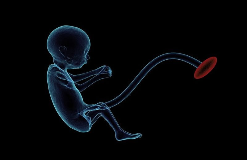
The placenta provides a supportive environment for developing mammalian fetuses. One of its most important roles is to supply nutrients, including amino acids, from the mother. But the molecular mechanisms behind the supply of amino acids are not well understood.
Now, a team led by Atsuo Ogura of the RIKEN BioResource Research Center has conducted a genetic study that explores the roles amino-acid transporters in the placenta play in development. Their major interest is cloning mammalian cells known as somatic cell nuclear transfer (SCNT), which offers tremendous potential for producing livestock with useful genetic traits, generating animal models for disease, and reproductive medicine.
“However, SCNT-generated embryos frequently show various developmental abnormalities, including an abnormally large placenta,” says Shogo Matoba, a senior research scientist in Ogura’s lab. “Only 1–5% of SCNT-cloned mouse embryos develop to term.”
A dragnet search for abnormally regulated genes in the placentas of these cloned embryos led the researchers to small neutral amino-acid transporter 4 (SNAT4), which belongs to a larger family of proteins that facilitate the delivery of a variety of amino acids. Ogura and Matoba made a series of genetic manipulations to better understand this protein’s importance in early development of mouse embryos.
After confirming that SNAT4 is specifically expressed in the healthy placenta, the researchers used the CRISPR–Cas9 gene-editing technology to generate mouse embryos that were deficient in SNAT4. The resulting pups were severely underweight, and only 28% were alive two weeks after delivery.
The researchers then bred SNAT-4-deficient males with normal females, and found that the resulting pregnancies still yielded severely underweight embryos with equally underdeveloped placentas. These changes were associated with reduced amino-acid levels in the circulation of fetal mice, hinting at a likely cause for this impaired development.
Since SNAT4 is strikingly overexpressed in SCNT-generated embryos, this could potentially explain the abnormally large placentas observed in this context.
These results also have implications for normal human reproduction. “The SNAT4-knockout mice showed phenotypes similar to those seen in intrauterine growth retardation,” says Matoba, referring to a developmental abnormality that can potentially lead to miscarriage. “These animals could be a valuable model for studying the mechanisms of this phenomenon in mammals—including humans.”
Matoba intends to investigate further how amino-acid transporters shape normal fetal development by knocking out other SNAT-encoding genes alongside SNAT4. He also wants to explore SNAT4’s function in non-placental tissues such as the liver.
Source:
RIKEN
Media Contacts:
Adam Philips – RIKEN
Image Source:
The image is in the public domain.
Original Research: Closed access
“Paternal knockout of Slc38a4/SNAT4 causes placental hypoplasia associated with intrauterine growth restriction in mice”. Shogo Matoba, Shoko Nakamuta, Kento Miura, Michiko Hirose, Hirosuke Shiura, Takashi Kohda, Nobuaki Nakamuta, and Atsuo Ogura.
PNAS doi:10.1073/pnas.1907884116.
Abstract
Paternal knockout of Slc38a4/SNAT4 causes placental hypoplasia associated with intrauterine growth restriction in mice
The placenta is critical in mammalian embryonic development because the embryo’s supply of nutrients, including amino acids, depends solely on mother-to-embryo transport through it. However, the molecular mechanisms underlying this amino acid supply are poorly understood. In this study, we focused on system A amino acid transporters Slc38a1/SNAT1, Slc38a2/SNAT2, and Slc38a4/SNAT4, which carry neutral, short-side-chain amino acids, to determine their involvement in placental or embryonic development. A triple-target CRISPR screen identified Slc38a4/SNAT4 as the critical amino acid transporter for placental development in mice. We established mouse lines from the CRISPR founders with large deletions in Slc38a4 and found that, consistent with the imprinted paternal expression of Slc38a4/SNAT4 in the placenta, paternal knockout (KO) but not maternal KO of Slc38a4/SNAT4 caused placental hypoplasia associated with reduced fetal weight. Immunostaining revealed that SNAT4 was widely expressed in differentiating cytotrophoblasts and maturing trophoblasts at the maternal–fetal interface. A blood metabolome analysis revealed that amino acid concentrations were globally reduced in Slc38a4/SNAT4 mutant embryos. These results indicated that SNAT4-mediated amino acid transport in mice plays a major role in placental and embryonic development. Given that expression of Slc38a4 in the placenta is conserved in other species, our Slc38a4/SNAT4 mutant mice could be a promising model for the analysis of placental defects leading to intrauterine growth restriction in mammals.