

Neuroscientists at Emory have refined a map showing which parts of the brain are activated during head rotation, resolving a decades-old puzzle. Their findings may help in the study of movement disorders affecting the head and neck, such as cervical dystonia and head tremor.
The results were published in Journal of Neuroscience.
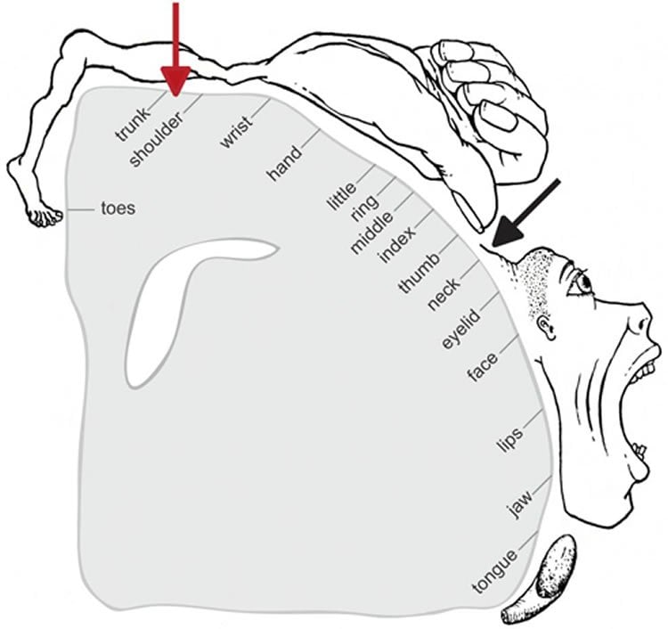
In landmark experiments published in the 1940s and 50s, Canadian neurosurgeon Wilder Penfield and colleagues determined which parts of the motor cortex controlled the movements of which parts of the body.
Penfield stimulated the brain with electricity in patients undergoing epilepsy surgery, and used the results to draw a “motor homunculus”: a distorted representation of the human body within the brain. Penfield assigned control of the neck muscles to a region between those that control the fingers and face, a finding inconsistent with some studies that came later.
Using modern functional MRI (magnetic resonance imaging), researchers at Emory University School of Medicine have shown that the neck’s motor control region in the brain is actually between the shoulders and trunk, a location that more closely matches the arrangement of the body itself.
“We can’t be that hard on Penfield, because the number of cases where he was able to study head movement was quite limited, and studying head motion as he did, by applying an electrode directly to the brain, creates some challenges,” says lead author Buz Jinnah, MD, professor of neurology, human genetics and pediatrics at Emory University School of Medicine.
The new location for the neck muscles makes more sense, because it corresponds to a similar map Penfield established of the sense of touch (the somatosensory cortex), Jinnah says.
Participants in brain imaging studies need to keep their heads still to provide accurate data, so volunteers were asked to perform isometric muscle contraction. They attempted to rotate their heads to the left or the right, even though head movement was restricted by foam padding and restraining straps.
First author Cecilia Prudente, a graduate student in neuroscience who is now a postdoctoral associate at the University of Minnesota, developed the isometric head movement task and obtained internal funding that allowed the study to proceed.
She and Jinnah knew that isometric exercises for the wrist activated the same regions of the motor cortex as wrist movements, and used that as a reference point in their study. During brain imaging, they were able to check that particular muscles were being tensed by directly monitoring volunteers’ muscles electronically.
When volunteers contracted their neck muscles, researchers were able to detect activation in other parts of the brain too, such as the cerebellum and the basal ganglia, which are known to be involved in movement control. This comes as no surprise, Jinnah says, since these regions also control movements of the hands and other body parts.
Prudente, Jinnah and colleagues have conducted a similar study with cervical dystonia patients, with the goal of comparing the patterns of brain activation between healthy volunteers and the patients. Cervical dystonia is a painful condition in which the neck muscles contract involuntarily and the head posture is distorted.
“These results may help guide future studies in humans and animals, as well as medical or surgical interventions for cervical dystonia and other disorders involving abnormal head movements,” Prudente says.
Emory co-authors: Randall Stilla, Cathrin Buetefisch, Shivangi Singh, Ellen Hess, Xiaoping Hu and Krish Sathian
Funding: The research was supported by the National Institute of Neurological Disorders and Stroke (U54NS065701, U54NS06571-03S1), the Office of Rare Diseases Research in the National Center for Advancing Translational Sciences, the Emory University Research Committee and the Emory Biomedical Imaging Technology Center.
Source: Quinn Eastman – Emory
Image Credit: Image courtesy of Journal of Neuroscience.
Original Research: Abstract for “Neural Substrates for Head Movements in Humans: A Functional Magnetic Resonance Imaging Study” by Cecilia N. Prudente, Randall Stilla, Cathrin M. Buetefisch, Shivangi Singh, Ellen J. Hess, Xiaoping Hu, Krish Sathian, and H.A. Jinnah in Journal of Neuroscience. Published online June 17 2015 doi:10.1523/JNEUROSCI.0851-15.2015
Abstract
Neural Substrates for Head Movements in Humans: A Functional Magnetic Resonance Imaging Study
The neural systems controlling head movements are not well delineated in humans. It is not clear whether the ipsilateral or contralateral primary motor cortex is involved in turning the head right or left. Furthermore, the exact location of the neck motor area in the somatotopic organization of the motor homunculus is still debated and evidence for contributions from other brain regions in humans is scarce. Because currently available neuroimaging methods are not generally suitable for mapping brain activation patterns during head movements, we conducted fMRI scans during isometric tasks of the head. During isometric tasks, muscle contractions occur without an actual movement and they have been used to delineate patterns of brain activity related to movements of other body parts such as the hands. Healthy individuals were scanned during isometric head rotation or wrist extension. Isometric wrist extension was examined as a positive control and to establish the relative locations of head and hand regions in the motor cortex. Electromyographic recordings of neck and hand muscles during scanning ensured compliance with the tasks. Increased brain activity during isometric head rotation was observed bilaterally in the precentral gyrus, both medial and lateral to the hand area, as well the supplementary motor area, insula, putamen, and cerebellum. These findings clarify the location of the neck region in the motor homunculus and help to reconcile some of the conflicting results obtained in earlier studies.
“Neural Substrates for Head Movements in Humans: A Functional Magnetic Resonance Imaging Study” by Cecilia N. Prudente, Randall Stilla, Cathrin M. Buetefisch, Shivangi Singh, Ellen J. Hess, Xiaoping Hu, Krish Sathian, and H.A. Jinnah in Journal of Neuroscience. Published online June 17 2015 doi:10.1523/JNEUROSCI.0851-15.2015


