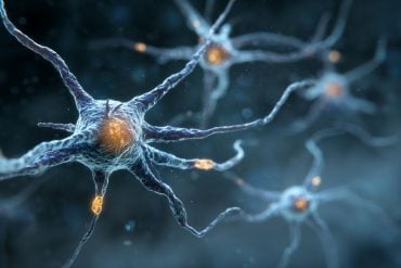
Summary: The brain needs constant energy to function, especially for managing neurotransmitter activity. A new study shows that when energy is depleted—such as during a stroke—neurons begin releasing glutamate in abnormal, self-amplifying bursts that can damage nerve cells.
Using a fluorescent sensor, researchers observed long-lasting, localized glutamate events that became more frequent under stress. These findings point to a dangerous feedback loop and highlight how disrupted metabolism leads to excitotoxicity, worsening outcomes in conditions like stroke.
Key Facts:
- Energy Crisis: Energy depletion halts normal glutamate release and triggers abnormal, self-reinforcing surges.
- Toxic Cascade: Excess extracellular glutamate harms neurons and impairs synaptic function.
- Intervention Clue: Blocking NMDA receptors significantly reduced harmful glutamate release events.
Source: RUB
Our brain requires a constant supply of energy. A disrupted energy supply, as it is for instance caused by a stroke, can have serious complications.
A team from the research group Cellular Neurobiology at Ruhr University Bochum, Germany, together with researchers from the Universities of Düsseldorf and Twente, investigated how an energy deficiency in the brain affects the release of the neurotransmitter glutamate.

They observed that under stress abnormal glutamate release events occur that self-amplify and that may thereby contribute to the damage of nerve cells.
The researchers, led by Dr. Tim Ziebarth, report their findings in the journal iScience on April 18, 2025.
Unusual glutamate releases occur more frequently upon energy depletion
Under normal conditions, brain tissue is sufficiently supplied with energy, which is in part required for the selective release and reuptake of neurotransmitters.
“However, if there is no longer enough energy available, this balance between neurotransmitter release and reuptake can quickly become disrupted,” Tim Ziebarth explains.
“Especially in the case of strokes, where the blood supply to the brain is interrupted, there is often an extracellular increase in the excitatory neurotransmitter glutamate, which severely impairs the function of synapses and the survival of the affected nerve cells.”
However, the underlying processes are only partially understood.
Using a model system, Tim Ziebarth observed a previously unknown, unconventional release mechanism that significantly increased the extracellular glutamate concentrations upon energy depletion.
For his measurements, he used a fluorescent sensor protein, which allowed him to visualize the release of glutamate in real-time. In addition to regular glutamate releases, which are typical for the synaptic activity of neurons, he also observed very unusual, local glutamate signals that were relatively large, long-lasting, and heterogeneous.
“Under normal conditions, these atypical events occurred only sporadically,” he reports.
“However, after inducing an energy deficiency, their frequency increased significantly.”
Normal glutamate release comes to a halt
Ultimately, these events were the main cause of the increase in extracellular glutamate concentration.
“It seems that under metabolic stress conditions, such as energy depletion, these atypical release events are particularly favored, which led to the accumulation of glutamate,” Professor Andreas Reiner concludes.
“In contrast, the normal glutamate release by neurons, which itself requires substantial amounts of energy, came to a halt.”
Similar observations had previously only been described for a migraine model.
In further experiments, the team was able to show that increased extracellular glutamate concentrations promoted additional release events. The process is therefore self-reinforcing. Conversely, the researchers were able to significantly reduce this type of glutamate release by inhibiting glutamate receptors, especially by inhibiting the subclass of NMDA receptors.
The study does not yet answer exactly how the unusual neurotransmitter releases occur and which cell types are responsible.
“Further investigations must also clarify how much this type of release actually contributes in stroke situations or in neurodegenerative diseases,” says Andreas Reiner.
However, it is well established that elevated glutamate concentrations are harmful to neurons.
About this neuroscience research news
Author: Julia Weiler
Source: RUB
Contact: Julia Weiler – RUB
Image: The image is credited to Neuroscience News
Original Research: Open access.
“Atypical Plume-Like Events Contribute to Glutamate Accumulation in Metabolic Stress Conditions” by Andreas Reiner et al. iScience
Abstract
Atypical Plume-Like Events Contribute to Glutamate Accumulation in Metabolic Stress Conditions
Neural glutamate homeostasis is important for health and disease. Ischemic conditions, like stroke, cause imbalances in glutamate release and uptake due to energy depletion and depolarization.
We here used the glutamate sensor SF-iGluSnFR(A184V) to probe how chemical ischemia affects the extracellular glutamate dynamics in slice cultures from mouse cortex.
SF-iGluSnFR imaging showed spontaneous glutamate release indicating synchronous network activity, similar to calcium imaging with GCaMP6f. Glutamate imaging further revealed local, atypically large, and long-lasting plume-like release events.
Plumes occurred with low frequency, independent of network activity, and persisted in tetrodotoxin (TTX).
Blocking glutamate uptake with TFB-TBOA favored plumes, whereas blocking ionotropic glutamate receptors (iGluRs) suppressed plumes.
During chemical ischemia plumes became more pronounced, overly abundant and contributed to large-scale glutamate accumulation.
Similar plumes were previously observed in cortical spreading depression and migraine models, and they may thus be a more general consequence of glutamate uptake dysfunctions in neurological and neurodegenerative diseases.


