Disturbances in the early stages of brain growth, such as preterm birth – when many of the brain’s structures have not yet fully developed – appears to affect the brain’s neuro-circuitry, which may explain premature babies’ higher risk of neurodevelopmental disorders including ADHD and autism spectrum disorder.
Researchers led by Natasha Lepore, PhD, of The Saban Research Institute of Children’s Hospital Los Angeles, have located significant alterations to specific surface regions of the brain. Described in a study published online this week by the journal Brain Structure and Function, their identification of neuroanatomical changes related to prematurity helps explain what brain structure and circuitry are affected, and may lead to designing effective prevention strategies and early interventional treatments for cognitive disabilities.
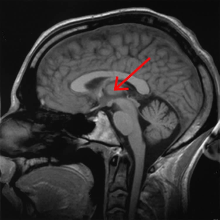
Using three-dimensional brain structural magnetic resonance imaging (MRI), Lepore and colleagues analyzed the structure and neural circuitry of two specific areas of the brain in 17 preterm and 19 term-born babies: the thalamus – the brain’s relay station, critical to sending and receiving sensory information – and the putamen, part of an intricate circuit connecting to the brain’s frontal lobe and involved in a number of different processes, most notably regulation of movement and learning.
While many studies have spotted alterations in various brain structures related to prematurity, this is the first study to link the structural abnormalities to specific neuro-circuitry, the communication pathways of the brain. To investigate these changes, the CHLA researchers performed a novel, combined analysis of the external shape and dimension of the surfaces of the thalamus and putamen, and compared the relative position of these structures to one another.
“We found that regional abnormalities of the thalamus are associated with alterations of the putamen, possibly due to disturbed development of shared frontal-subcortical connectivity,” said first author Yi Lao, MS, of the Department of Radiology at CHLA. More specifically, she added, the significantly correlated regions in these two structures point to frontal and sub-cortical pathways that are essential to important functions such as attention, decision-making, planning, abstract reasoning and memory.
Lepore adds that, for the first time, they have demonstrated the feasibility of using measurements of these abnormalities in the brain of preterm newborns as potential indicators of risk for future cognitive and behavioral problems.
“The ability to identify structural signs of neurodevelopmental disease shortly after birth in premature infants could allow for early interventions, increasing the child’s social and learning behaviors as they age,” said Lepore.
Additional contributors include Yalin Wang, PhD, and Jie Shi, MS, Arizona State University; Rafael Ceschin, MS, Children’s Hospital of Pittsburgh; Ashok Panigrahy, MD, Children’s Hospital Los Angeles and Children’s Hospital of Pittsburgh; and Marvin D. Nelson, MD, Children’s Hospital Los Angeles.
This work was supported by the National Institutes of Health through NIH grant 5K23-NS063371 and grants R21EB012177 and R21AG043760.
Contact: Debra Kain – Children’s Hospital Los Angeles
Source: Children’s Hospital Los Angeles press release
Image Source: The image is credited to AxelBoldt and is licensed Creative Commons Attribution-ShareAlike 3.0 Unported
Original Research: Abstract for “Thalamic alterations in preterm neonates and their relation to ventral striatum disturbances revealed by a combined shape and pose analysis” by Yi Lao, Yalin Wang, Jie Shi, Rafael Ceschin, Marvin D. Nelson, Ashok Panigrahy, and Natasha Leporé in Brain Structure and Function. Published online November 1 2014 doi:10.1007/s00429-014-0921-7