Summary: Researchers developed a groundbreaking technology to track when brain cells deactivate, a key process known as inhibition. This technique allows a deeper understanding of normal brain functions and disorders like depression, PTSD, and Alzheimer’s.
By using optogenetics and identifying changes in the pyruvate dehydrogenase (PDH) protein, the team can now observe how the brain conserves energy by rapidly shutting off this protein post-neuron firing. This discovery opens new avenues for studying brain cell inhibition and its role in various diseases.
Key Facts:
- The new technology tracks the deactivation of neurons, shedding light on how the brain regulates activity through inhibition.
- The study revealed rapid changes in the PDH protein, key to energy production, immediately after brain cells stop firing.
- This technique can be instrumental in understanding and potentially treating a range of brain disorders and metabolic diseases.
Source: Scripps Research Institute
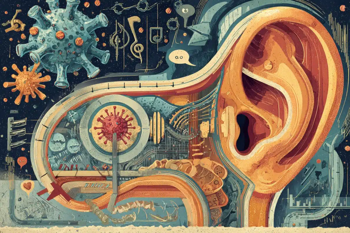
For decades, scientists have studied the intricate activity patterns in human and animal brains by observing when different groups of brain cells turn on. Equally important to understanding the brain and related diseases, however, is knowing how long those neurons stay active and when they turn off again.
Now, scientists at Scripps Research have developed a new technology that lets them track when, after a burst of activity, brain cells shut off—a process known as inhibition.

The technique, published in Neuron on January 23, 2024, provides a new way to study not only the normal functioning of the brain, but how the brain’s “off switches” may go awry in normal behaviors as well as in diseases and disorders, including depression, post-traumatic stress disorder and Alzheimer’s disease.
“It’s generally agreed that the inhibition, of neurons is really the major way the brain is regulating activity,” says senior author Li Ye, Ph.D., professor and Abide-Vividion Chair at Scripps Research. “Scientists have been looking for a way to look at inhibition on a more trackable way, and until now, few had found it.”
For pioneering the new approach, Ye teamed up with John Yates, a professor of Molecular Medicine at Scripps Research. They wanted to study how brain cells changed when they were actively firing—emitting an electrical charge to pass messages to their neighbors—compared to when they were done firing.
The scientists used optogenetics, in which cells’ activity can be controlled using light, to repeatedly activate and inhibit the cells. Then, they measured levels and characteristics of different proteins and their modifications. They identified that one protein, pyruvate dehydrogenase (PDH), was very rapidly changed immediately after brain cells were inhibited.
“When neurons are firing, you need a lot of energy, and this PDH protein is involved in producing that energy,” explains Ye. “But the brain really wants to conserve energy, so when a cell is done firing, we found that the brain rapidly shuts off the PDH protein. This happened much faster than anything else we saw in gene expression.”
To shut off PDH, the researchers found, cells add molecular tags called phosphates to the protein. Ye and his colleagues found antibodies that only recognized this inactive, phosphorylated form of PDH (pPDH). To test whether levels of phosphorylated PDH (or pPDH) could be used as a proxy for brain cell inhibition,
Ye’s team used these antibodies to measure pPDH in mice that had been given anesthesia. Nearly the entire brain lit up with high levels of pPDH, correctly showing how most of the brain is inactive during anesthesia.
The group also studied levels of pPDH when animals were exposed to bright light that was then shut off. Brain cells in the visual cortex, responsible for vision, had low levels of pPDH when being exposed to light (because the active form of PDH would be required to give these cells signaling energy), but high levels of phosphorylated protein immediately increased after the light was off.
Ye’s group also used the new technique to study a less understood process: how the brain turns off the feeling of hunger after a meal. They showed how brain cells related to appetite shut off when a mouse starts to eat.
Those findings could have implications for better understanding appetite, obesity and some weight loss drugs. More broadly, the pPDH antibodies could be used to compare levels of brain cell inhibition in healthy people and those with a variety of brain and metabolic diseases.
“There are a lot of questions that this technology can help us answer,” says Ye. “If the brain can’t turn cells off, or if they’re turned off faster or slower than usual, what happens? How does the inhibition of neurons play a role in different diseases?”
Ye and his colleagues are continuing to fine-tune the use of pPDH, but they say that other researchers are already using the technology—the antibodies used to measure pPDH are commercially available.
In addition to Ye and Yates, authors of the study, “Phosphorylation of pyruvate dehydrogenase inversely associates with neuronal activity,” include Dong Yang, Yu Wang, Tianbo Qi, Xi Zhang, Leyao Shen, Jingrui Ma, Zhengyuan Pang, Neeraj K. Lal, Daniel B. McClatchy, Saba Heydari Seradj, Verina H. Leung, Kristina Wang, Yi Xie, Filip S. Polli, Anton Maximov, Hollis T. Cline and Vineet Augustine of Scripps Research; and Oscar Christian Gonzalez and Luis de Lecea of Stanford University.
Funding: This work was supported by funding from the National Institutes of Health (DP2DK128800), and BRAIN initiative/NIMH (MH132570and the Dorris Scholar Award.
About this neurotech and neuroscience research news
Author: Scripps Research Communications Office
Source: Scripps Research Institute
Contact: Scripps Research Communications Office – Scripps Research Institute
Image: The image is credited to Neuroscience News
Original Research: Open access.
“Phosphorylation of pyruvate dehydrogenase inversely associates with neuronal activity” by Li Ye et al. Neuron
Abstract
Phosphorylation of pyruvate dehydrogenase inversely associates with neuronal activity
Highlights
- Establishing an optogenetic-based quantitative proteomic screening platform
- Unbiased screening identified that neural activity decreases pPDH
- pPDH level negatively correlates with neural activity in vitro and in vivo
- pPDH as a novel inverse activity marker (IAM) to track reduced neuronal activity
Summary
For decades, the expression of immediate early genes (IEGs) such as FOS has been the most widely used molecular marker representing neuronal activation. However, to date, there is no equivalent surrogate available for the decrease of neuronal activity.
Here, we developed an optogenetic-based biochemical screen in which population neural activities can be controlled by light with single action potential precision, followed by unbiased phosphoproteomic profiling.
We identified that the phosphorylation of pyruvate dehydrogenase (pPDH) inversely correlated with the intensity of action potential firing in primary neurons.
In in vivo mouse models, monoclonal antibody-based pPDH immunostaining detected activity decreases across the brain, which were induced by a wide range of factors including general anesthesia, chemogenetic inhibition, sensory experiences, and natural behaviors.
Thus, as an inverse activity marker (IAM) in vivo, pPDH can be used together with IEGs or other cell-type markers to profile and identify bi-directional neural dynamics induced by experiences or behaviors.