

Summary: A new study reports two weeks following a concussion, myelin surrounding neurons appear to loosen. However, the loosening returns to normal two months after a concussion, researchers say.
Source: University of British Columbia.
Detailed scans of concussed university hockey players found that the protective fatty tissue surrounding brain cell fibers was loosened two weeks after the injury–even though the athletes felt fine and were deemed ready to return to the ice.
A loosening of that insulation, called myelin, slows the transmission of electrical signals between brain cells, or neurons. Researchers have previously shown in animals that this loosened myelin can completely deteriorate with subsequent blows–a condition that resembles the neurodegenerative disease multiple sclerosis.
“This is the first solid evidence in humans that concussions loosen myelin,” said Alex Rauscher, an Associate Professor in the Department of Pediatrics and the Canada Research Chair in Developmental Neuroimaging at the University of British Columbia. “And it was detected two weeks after the concussion, when the players said they felt fine and were deemed ready to play through standard return-to-play evaluations. So athletes may be returning to play sooner than they should.”
Published this month in Frontiers in Neurology, this is the third study arising from the unusual before-and-after study of UBC hockey players. The 45 athletes had their brains scanned with magnetic resonance imaging (MRI) before the season began; if they were concussed, they were re-scanned three days afterwards, two weeks afterwards, and two months afterwards. Eleven athletes were concussed during the season, and most of them underwent the additional MRI scans.
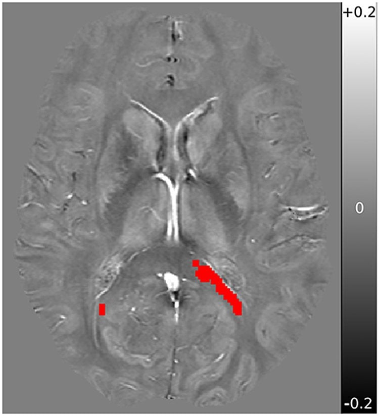
Conventional MRI imaging–the kind done in hospitals to assess brain injury–does not reveal myelin loosening. Rauscher and postdoctoral research fellow Alex Weber used advanced digital analysis of the scans, using a UBC-developed, pixel-based statistical analysis to find changes that visual inspection could not reveal.
Previous analysis of the concussed athletes’ scans, published by Rauscher in 2016, had shown changes to the myelin in the corpus callosum–the part of the brain that helps the brain’s two hemispheres communicate, and is most susceptible to damage from sudden collisions against the interior of the skull. But the researchers didn’t know whether the myelin was diminished, akin to multiple sclerosis, or altered in some other way.
The good news is that it was something else–a temporary loosening around the nerve fibers that connect brain cells. When the concussed players were re-scanned two months after their concussions, their myelin had returned to normal.
But Weber says the findings provide a convincing reason to keep concussed athletes on the bench even if they no longer exhibit any symptoms, as measured by a standard test of cognitive abilities, balance, coordination and mood.
“These results show that there is some damage happening below the surface at least two weeks after a concussion,” Weber says. “Passing a concussion test may not be a reliable indicator of whether their brain has truly healed. We might need to build in more waiting time to prevent any long-term damage.”
Source: Brian Kladko – University of British Columbia
Publisher: Organized by NeuroscienceNews.com.
Image Source: NeuroscienceNews.com image is credited to credited to Webber et al.
Original Research: Open access research for “Pathological Insights From Quantitative Susceptibility Mapping and Diffusion Tensor Imaging in Ice Hockey Players Pre and Post-concussion” by Alexander M. Weber, Anna Pukropski, Christian Kames, Michael Jarrett, Shiroy Dadachanji, Jack Taunton, David K. B. Li and Alexander Rauscher in Frontiers in Neuroscience. Published August 6 2018.
doi:10.3389/fneur.2018.00575
[cbtabs][cbtab title=”MLA”]University of British Columbia”Concussion Loosens Insulation Around Brain Cells.” NeuroscienceNews. NeuroscienceNews, 4 September 2018.
<https://neurosciencenews.com/myelin-concussion-9794/>.[/cbtab][cbtab title=”APA”]University of British Columbia(2018, September 4). Concussion Loosens Insulation Around Brain Cells. NeuroscienceNews. Retrieved September 4 2018 from https://neurosciencenews.com/myelin-concussion-9794/[/cbtab][cbtab title=”Chicago”]University of British Columbia”Concussion Loosens Insulation Around Brain Cells.” https://neurosciencenews.com/myelin-concussion-9794/ (accessed September 4 2018).[/cbtab][/cbtabs]
Abstract
Pathological Insights From Quantitative Susceptibility Mapping and Diffusion Tensor Imaging in Ice Hockey Players Pre and Post-concussion
Myelin sensitive MRI techniques, such as diffusion tensor imaging and myelin water imaging, have previously been used to reveal changes in myelin after sports-related concussions. What is not clear from these studies, however, is how myelin is affected: whether it becomes degraded and possibly removed, or whether the myelin sheath loosens and becomes “decompacted”. Previously, our team revealed myelin specific changes in ice hockey players 2 weeks post-concussion using myelin water imaging. In that study, 45 subjects underwent a pre-season baseline scan, 11 of which sustained a concussion during play and received follow-up scans: eight were scanned within 3 days, 10 were scanned at 14 days, and nine were scanned at 60 days. In the current retrospective analysis, we used quantitative susceptibility mapping, along with the diffusion tensor imaging measures axial diffusivity and radial diffusivity, to investigate this myelin disruption. If sports-related concussive hits lead to myelin fragmentation in regions of lowered MWF, this should result in a measurable increase in magnetic susceptibility, due to the anisotropic myelin fragmenting into isotropic myelin debris, and the diamagnetic myelin tissue being removed, while no such changes should be expected if the myelin sheath simply loosens and becomes decompacted. An increase in radial diffusivity would likewise reveal myelin fragmentation, as myelin sheaths block water diffusion out of the axon, with little to no changes expected for myelin sheath loosening. Statistical analysis of the same voxels-of-interest that were found to have reduced myelin water fraction 2 weeks post-concussion, revealed no statistically significant changes in magnetic susceptibility, axial diffusivity, or radial diffusivity at any time-point post-concussion. This suggests that myelin water fraction changes are likely due to a loosening of the myelin sheath structure, as opposed to fragmentation and removal of myelin debris.


