

Summary:
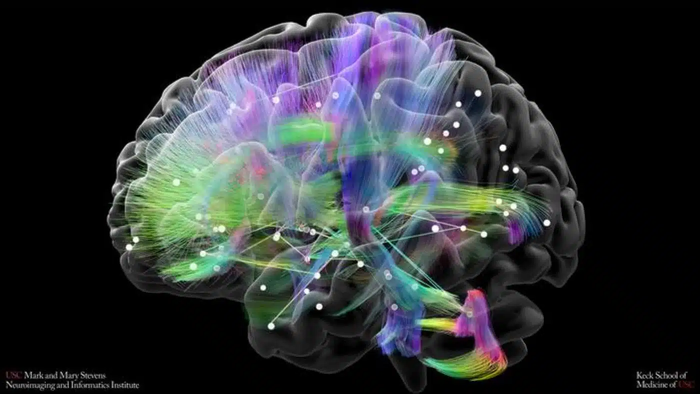
A multi-institutional consortium led by researchers at the Keck School of Medicine of USC’s Stevens Neuroimaging and Informatics Institute, Stanford University, and Johns Hopkins University has received a $4.5 million grant from BD² to investigate the biological drivers of bipolar mood switching. Focusing on a specialized population of neurons termed “retrosplenial switch cells,” the team will combine postmortem single-cell histology, polygenic risk scores from a 4,000-patient clinical cohort, and large-scale ENIGMA connectome neuroimaging to decipher how the retrosplenial cortex alternates between depression and mania.
Key Facts:
- Focus on “Switch Cells”: Building on recent discoveries implicating the retrosplenial cortex in psychiatric illness, researchers hypothesize that specialized retrosplenial neurons act as master biological gates regulating transitions between manic and depressive poles.
- $4.5 Million BD² Discovery Grant: The three-year initiative is supported by Breakthrough Discoveries for thriving with Bipolar Disorder (BD²), uniting investigators at USC Stevens INI, Stanford, and Johns Hopkins.
- Multi-Scale Triangulation: The project connects three distinct scientific layers: single-cell molecular profiling of postmortem human brain tissue, polygenic prediction modeling within the 4,000-participant BD² Integrated Network, and macroscale structural/functional connectomics via the global ENIGMA consortium.
Source: Keck School of Medicine of USC
Bipolar disorder affects nearly 3% of the global population, characterized by unpredictable, cyclic oscillations between debilitating depressive lows and elevated, high-energy manic or hypomanic states. Despite its prevalence and severity, the fundamental neurobiological mechanisms that govern mood switching, the physical biological process that shifts the brain from one extreme affective state to another, remain one of the most stubborn mysteries in clinical psychiatry.
As a result, current treatment regimens often rely on broad mood stabilizers and trial-and-error pharmacology, leaving many patients struggling with chronic instability, medication side effects, or misdiagnoses.
To resolve this biological blind spot, a cross-disciplinary team led by Christopher R.K. Ching, PhD, assistant professor of research neurology at the Mark and Mary Stevens Neuroimaging and Informatics Institute (Stevens INI) at the Keck School of Medicine of USC, has launched a multi-scale investigation. Co-led by Laramie Duncan, PhD, of Stanford University, and Fernando Goes, MD, of Johns Hopkins University, the project is backed by a $4.5 million, three-year Discovery Research grant from Breakthrough Discoveries for thriving with Bipolar Disorder (BD²).
The Retrosplenial Cortex and Its “Switch Cells”
Located in the posterior medial cortex, the retrosplenial cortex (RSC) has historically been studied for its structural role in episodic memory retrieval, spatial orientation, and navigation. When compromised by focal lesions or pathological insults, damage to the region frequently produces severe dissociative phenomena—states where individuals feel profoundly detached from their personal identity, emotional reality, or external surroundings.
In a landmark 2025 study published in Nature Neuroscience, Duncan and colleagues revealed that specific neuronal subtypes within the retrosplenial cortex show pronounced transcriptional and genetic vulnerability in bipolar disorder.
The researchers dubbed this distinct population “retrosplenial switch cells,” hypothesizing that these cells serve as critical regulatory nodes in the brain’s mood architecture. Under normal conditions, they maintain affective stability; when genetically or functionally disrupted, their breakdown may trigger the switch into mania or depression.
Connecting Single Cells to Global Brain Networks
The new research initiative bridges three complementary biological dimensions:
- Molecular and Cellular Profiling: Using high-resolution molecular labeling and single-cell spatial transcriptomics, the investigators will map cell populations within the retrosplenial cortex of postmortem brain tissue donated by individuals with and without bipolar disorder. This will pinpoint exact cellular anomalies and morphological variances unique to the condition.
- Genomic Risk and Clinical Trajectories: Leveraging the BD² Integrated Network, a longitudinal cohort enrolling 4,000 individuals diagnosed with bipolar disorder, the team will evaluate whether composite polygenic risk scores (PRS) can reliably predict the frequency of mood switching episodes and individual responses to standard mood-stabilizing therapeutics.
- Macroscale Connectome Neuroimaging: The researchers will analyze structural and functional magnetic resonance imaging (fMRI) data across the BD² cohort to chart how the retrosplenial cortex communicates with distant cortical, limbic, and subcortical networks.
“We will apply some of the cutting-edge structural and functional brain network modeling approaches we have developed in our large, international ENIGMA Bipolar group studies,” said Melody Kang, a USC Neuroscience PhD student working on the project. Ching directs the ENIGMA Bipolar Disorder Working Group, a worldwide collaborative network pooling neuroimaging and clinical datasets across more than 250 institutions worldwide.
A Multidisciplinary Discovery Engine
By evaluating the retrosplenial cortex across genomic, cellular, and whole-brain network scales, the team aims to establish an integrated model of bipolar mood instability.
“This is the type of ambitious, multidisciplinary project that would have been difficult to fund through traditional scientific grants,” Ching noted. “This BD2 project gives us a rare opportunity to link the changes we observe in the retrosplenial cortex at the human neuroimaging level to the underlying cellular and genetic factors that may lead to more clinically actionable markers aimed at improving diagnosis and treatment of bipolar disorder.”
Arthur W. Toga, PhD, director of the Stevens INI, highlighted the collaborative framework: “Mood switching shapes daily life for people with bipolar disorder, yet its biology remains poorly understood. This project brings imaging, genetics, and cell biology together to address a single question, using data openly shared across research teams. That combination gives us a much better chance of finding answers that matter to patients.”
While exploratory discovery research that does not yet test direct pharmacological compounds, the findings aim to identify specific molecular pathways within retrosplenial switch cells, laying the groundwork for targeted, next-generation therapeutics engineered to arrest the mood-switching cycle.
Editorial Notes:
- This article was edited by a Neuroscience News editor.
- Journal paper will be reviewed in full upon release.
- Additional context added by our staff.
About this neuroscience Research:
- Media Contact: Laura LeBlanc
- Source: USC
- Image Credit: Image credited to Stevens INI


