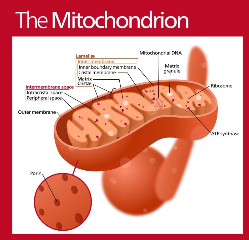
Researchers at the UC San Diego School of Medicine have demonstrated for the first time that veterans of the 1990-91 Persian Gulf War who suffer from “Gulf War illness” have impaired function of mitochondria – the energy powerhouses of cells.
The findings, published in the March 27, 2014 issue of PLOS ONE, could help lead to new treatments benefitting affected individuals – and to new ways of protecting servicepersons (and civilians) from similar problems in the future, said principal investigator Beatrice A. Golomb MD, PhD, professor of medicine.
Golomb, with associate Hayley Koslik and Gavin Hamilton, PhD, a research scientist and magnetic resonance physicist, used the imaging technology to compare Gulf War veterans with diagnosed Gulf War illness to healthy controls. Cases were matched by age, sex and ethnicity.
The technique used – 31-phosphorus magnetic resonance spectroscopy or 31P-MRS – reveals amounts of phosphorus-containing compounds in cells. Such compounds are important for cell energy production, in particular phosphocreatine or PCr, which declines in muscle cells during exercise. PCr recovery takes longer when mitochondrial function is impaired, and delayed recovery is recognized as a robust marker of mitochondrial dysfunction.

Affected Gulf War veterans displayed significantly delayed PCr recovery after an exercise challenge. In fact, said Golomb, there was almost no overlap in the recovery times of Gulf War illness veterans compared to controls: All but one control participant had a recovery time-constant clustered under 31 seconds. In contrast, all but one Gulf Illness veteran had a recovery time-constant exceeding 35 seconds, with times ranging as high as 70 seconds.
There were 14 participants in the study: seven Gulf War illness cases and seven matching controls. Golomb notes that the use of 1:1 matching markedly improves statistical “power,” allowing a smaller sample size. The separation between the two groups was “visibly striking, and the large average difference was statistically significant,” she said.
Golomb noted that impaired mitochondrial function accounts for numerous features of Gulf War illness, including symptoms that have been viewed as perplexing or paradoxical.
“The classic presentation for mitochondrial illness involves multiple symptoms spanning many domains, similar to what we see in Gulf War illness. These classically include fatigue, cognitive and other brain-related challenges, muscle problems and exercise intolerance, with neurological and gastrointestinal problems also common.”
There are other similarities between patients with mitochondrial dysfunction and those suffering from Gulf War illness: Additional symptoms appear in smaller subsets of patients; varying patterns of symptoms and severity among individuals; different latency periods across symptoms, or times when symptoms first appear; routine blood tests that appear normal.
“Some have sought to ascribe Gulf War illness to stress,” said Golomb, “but stress has proven not to be an independent predictor of the condition. On the other hand, Gulf veterans are known to have been widely exposed to acetylcholinesterase inhibitors, a chemical class found in organophosphate and carbamate pesticides, nerve gas and nerve gas pre-treatment pills given to troops.
“These inhibitors have known mitochondrial toxicity and generally show the strongest and most consistent relationship to predicting Gulf War illness. Mitochondrial problems account for which exposures relate to Gulf War illness, which symptoms predominate, how Gulf War illness symptoms manifest themselves, what objective tests have been altered, and why routine blood tests have not been useful.”
Funding for this research came, in part, from a UC San Diego Academic Senate Award and the U.S. Department of Defense.
Contact: Scott LaFee – UCSD
Source: UCSD press release
Image Source: The image is credited Kelvinsong. The author has released the image into the public domain.
Original Research: Full open access research for “Mitochondrial Dysfunction in Gulf War Illness Revealed by 31Phosphorus Magnetic Resonance Spectroscopy: A Case-Control Study” by Hayley J. Koslik, Gavin Hamilton, and Beatrice A. Golomb in PLOS ONE. Published online March 27 2014 doi:10.1371/journal.pone.0092887
Mitochondrial Dysfunction in Gulf War Illness Revealed by 31Phosphorus Magnetic Resonance Spectroscopy: A Case-Control Study
Approximately 1/3 of 1990-1 Gulf War veterans developed chronic multisymptom health problems. Implicated exposures bear mechanisms that adversely affect mitochondria. Symptoms emphasize fatigue, cognition and muscle (brain and muscle are aerobically demanding); with protean additional domains affected, compatible with mitochondrial impairment. Recent evidence supports treatments targeting cell bioenergetics (coenzyme10) to benefit Gulf War illness symptoms. However, no evidence has directly documented mitochondrial or bioenergetic impairment in Gulf War illness.
“Mitochondrial Dysfunction in Gulf War Illness Revealed by 31Phosphorus Magnetic Resonance Spectroscopy: A Case-Control Study” by Hayley J. Koslik, Gavin Hamilton, and Beatrice A. Golomb in PLOS ONE, March 27 2014 doi:10.1371/journal.pone.0092887


