Summary: Study reveals the detailed internal anatomy of mini-brains for the first time.
Source: Wyss Center for Bio and Neuroengineering
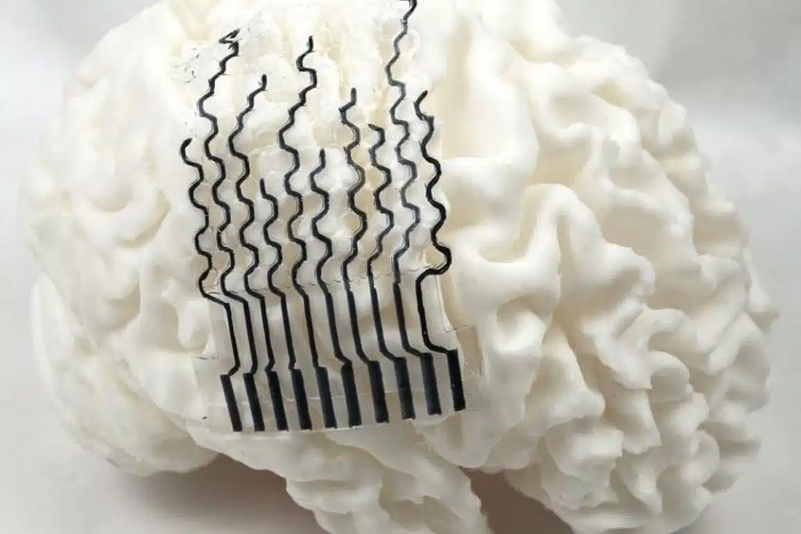
‘Mini-brains’ are pin-head sized collections of several different types of human brain cell. They are used as a tool, allowing scientists to learn about how the brain develops, study disease and test new medicines. Personalized ‘mini-brains’ can be grown from stem cells generated from a sample of human hair or skin and could shed light on how brain disease progresses in an individual and how this person may respond to drugs.
Research published today by a team of scientists and engineers from HEPIA and the Wyss Center for Bio and Neuroengineering, in the journal Frontiers in Bioengineering and Biotechnology, has revealed the detailed internal anatomy of ‘mini-brains’, for the first time.
“Despite advances in growing ‘mini-brains’, it has been difficult to understand in detail what is going on inside – until now,” said Professor Adrien Roux from the Tissue Engineering Laboratory, HEPIA, senior author on the paper.
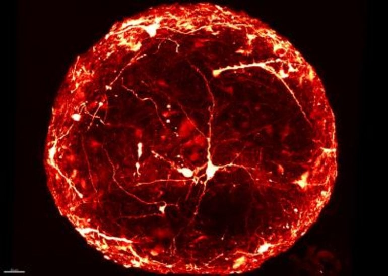
“Typically, to look inside a ‘mini-brain’, we slice it thinly and view it on a slide under a microscope. This is a slow process that can damage the sample. Now, for the first time, we have produced high resolution 3D images of single neurons within intact ‘mini-brains’, revealing their remarkable complexity,” added Dr Subashika Govindan, lead author on the paper, who carried out the work at HEPIA and is now Wellcome DBT early career fellow at the Indian Institute of Technology Madras (IITM).
The team combined a novel technique for labeling individual neurons with a method to make the whole sample completely transparent.
Leveraging the Wyss Center’s microscopy capabilities, the team developed a state-of-the-art custom module, including a bespoke sample holder and sensitive imaging detectors, for capturing 3D images of entire intact ‘mini-brains’, without slicing them. They were then able to visualize and analyze the 3D morphology of specific neurons and their anatomical distribution inside the ‘mini-brains’.
Dr Laura Batti, Microscopy Facility Manager at the Wyss Center said: “Human ‘mini-brains’ have a life span of more than a year and, with our new ability to visualize them in more detail, we can envision benefits such as reducing some animal testing.”
The new approach could also enable imaging of large numbers of ‘mini-brains’, making it suitable for high-throughput screening for drug discovery or toxicity testing. It is reproducible and cost-effective and could potentially help accelerate personalized medicine studies.
About this brain organoid research news
Source: Wyss Center for Bio and Neuroengineering
Contact: Jo Bowler – Wyss Center for Bio and Neuroengineering
Image: The image is credited to HEPIA
Original Research: Open access.
“Mass Generation, Neuron Labeling, and 3D Imaging of Minibrains” by Subashika Govindan et al. Frontiers in Bioengineering and Biotechnology
Abstract
Mass Generation, Neuron Labeling, and 3D Imaging of Minibrains
Minibrain is a 3D brain in vitro spheroid model, composed of a mixed population of neurons and glial cells, generated from human iPSC derived neural stem cells. Despite the advances in human 3D in vitro models such as aggregates, spheroids and organoids, there is a lack of labeling and imaging methodologies to characterize these models. In this study, we present a step-by-step methodology to generate human minibrain nurseries and novel strategies to subsequently label projection neurons, perform immunohistochemistry and 3D imaging of the minibrains at large multiplexable scales. To visualize projection neurons, we adapt viral transduction and to visualize the organization of cell types we implement immunohistochemistry. To facilitate 3D imaging of minibrains, we present here pipelines and accessories for one step mounting and clearing suitable for confocal microscopy. The pipelines are specifically designed in such a way that the assays can be multiplexed with ease for large-scale screenings using minibrains and other organoid models. Using the pipeline, we present (i) dendrite morphometric properties obtained from 3D neuron morphology reconstructions, (ii) diversity in neuron morphology, and (iii) quantified distribution of progenitors and POU3F2 positive neurons in human minibrains.