

Summary: Researchers identified several brain areas that acted as hubs for information processing across brain networks that contribute to memory recall. They observed how activation patterns within these networks differed on an individual level, based on personal levels of recall detail and imagination.
Source: University of Rochester
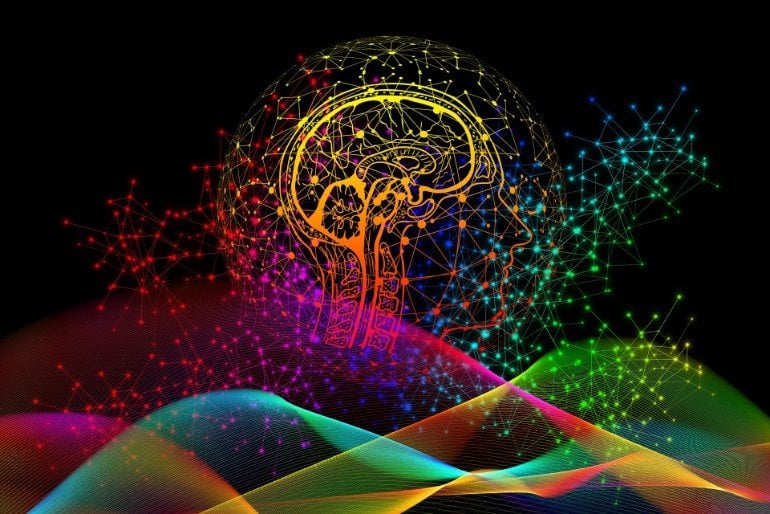
While the broad architecture and organization of the human brain is universal, new research shows how the differences between how people reimagine common scenarios can be observed in brain activity and quantified. These unique neurological signatures could ultimately be used to understand, study, and even improve treatment of disorders such as Alzheimer’s disease.
“When people imagine similar types of events, each person does it differently because they have different experiences,” said Feng (Vankee) Lin, Ph.D., R.N. “Our research demonstrates that we can decode the complex information in the human brain related to everyday life and identify neural ‘fingerprints’ that are unique to each individual’s remembered experience.” Lin is an associate professor in the University of Rochester Del Monte Institute for Neuroscience and co-author of the study which appears in the journal Nature Communications.
In the study, researchers asked 26 participants to recall common scenarios, such as driving, attending a wedding, or eating out at a restaurant. The scenarios were broad enough so that each participant would reimagine them differently. For example, when researchers asked volunteers to vividly remember and describe an occasion involving dancing, one person might recall watching their daughter participating in a dance recital, while another may imaging themselves dancing at a Bar Mitzvah.
The participant’s verbal descriptions were mapped to a computational linguistic model that approximates the meaning of the words and creates numerical representation of the context of the description. They were also asked to rate aspects of the remembered experience, such as how strongly it was associated with sound, color, movement, and different emotions.
The study volunteers were then placed in a functional MRI (fMRI) and asked to reimagine the experience while researchers measured which areas of the brain were activated. Using the fMRI data and the subject’s verbal descriptions and ratings, researchers were able to isolate brain activity patterns associated with that individual’s experiences. For instance, if the participant imagined driving through a red light in the scenario, areas of the brain associated with recalling motion and color would be activated. Using this data, the researchers built a functional model of each participant’s brain, essentially creating a unique signature of their neurological activity.
The researchers were able to identify several areas of the brain that served as hubs for processing information across brain networks that contribute to recalling information about people, objects, places, emotions, and sensations. The team was also able to observe how activation patterns within these networks differed on an individual level depending upon the details of each person’s recollections and imagination.
“One of the goals of cognitive science is to understand how memories are represented and manipulated by the human brain,” said Andrew Anderson, Ph.D., with the Del Monte Institute for Neuroscience and co-author of the study. “This study shows that fMRI can measure brain activity with sufficient signal to identify meaningful interpersonal differences in the neural representation of complex imagined events that reflect each individual’s unique experience.”
In addition to expanding our understanding of how the brain is networked, the authors point out that many of the key regions they identified tend to decline in function as we age and are vulnerable to the degeneration that occurs in disease like Alzheimer’s. The findings could lead to new ways to diagnose and study disorders associated with irregular memory deficits, including dementia, schizophrenia, and depression, and perhaps even personalize treatments and predict which therapies will be more effective.
Additional co-authors include Kelsey McDermott, Brian Rooks, Kathi Heffner, and David Dodell-Feder with the University of Rochester.
Funding: The study was funded with support from the National Center for Advancing Translational Sciences of the National Institutes of Health and the URMC Clinical the Translational Science Institute.
About this memory research news
Source: University of Rochester
Contact: Mark Michaud – University of Rochester
Image: The image is in the public domain
Original Research: Open access.
“Decoding individual identity from brain activity elicited in imagining common experiences” by Andrew James Anderson, Kelsey McDermott, Brian Rooks, Kathi L. Heffner, David Dodell-Feder & Feng V. Lin. Nature Communications
Abstract
Decoding individual identity from brain activity elicited in imagining common experiences
Everyone experiences common events differently. This leads to personal memories that presumably provide neural signatures of individual identity when events are reimagined. We present initial evidence that these signatures can be read from brain activity. To do this, we progress beyond previous work that has deployed generic group-level computational semantic models to distinguish between neural representations of different events, but not revealed interpersonal differences in event representations. We scanned 26 participants’ brain activity using functional Magnetic Resonance Imaging as they vividly imagined themselves personally experiencing 20 common scenarios (e.g., dancing, shopping, wedding). Rather than adopting a one-size-fits-all approach to generically model scenarios, we constructed personal models from participants’ verbal descriptions and self-ratings of sensory/motor/cognitive/spatiotemporal and emotional characteristics of the imagined experiences. We demonstrate that participants’ neural representations are better predicted by their own models than other peoples’. This showcases how neuroimaging and personalized models can quantify individual-differences in imagined experiences.


