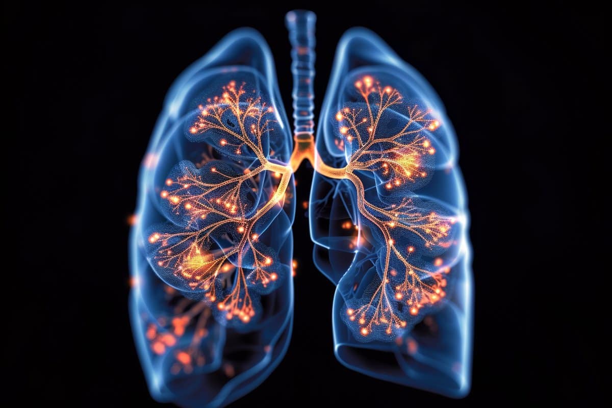
Summary: Researchers unveiled a groundbreaking mechanism where the lungs directly inform the brain of infections, altering traditional views on sickness response. This study, conducted in mice, demonstrates that neurological pathways, rather than just immune responses, are responsible for symptoms of illness.
The findings suggest that treating respiratory infections and chronic lung conditions might require approaches that target both the nervous system and the pathogen.
Additionally, the study observed a gender difference in sickness behavior, potentially offering a scientific basis for the so-called “man flu,” with males showing greater dependence on neuronal communications during illness.
Key Facts:
- Direct Lung-Brain Communication: The lungs use neurons involved in the pain pathway to alert the brain about infections, leading to symptoms of sickness through nervous system activation rather than solely through the immune response.
- Implications for Treatment: Understanding this lung-brain dialogue opens possibilities for dual treatment strategies that address both the infection and its neurological impact, offering hope for better management of respiratory conditions.
- Gender Differences in Sickness Response: The study found male mice showed more severe sickness behaviors than females under the same conditions, suggesting neuronal communication plays a more significant role in males, shedding light on gender disparities in illness experiences.
Source: University of Calgary
University of Calgary researchers have discovered the lungs communicate directly with the brain when there is an infection. Findings show the brain plays a critical role in triggering the symptoms of sickness, which may change the way we treat respiratory infections and chronic conditions.
“The lungs are using the same sensors and neurons in the pain pathway to let the brain know there’s an infection,” says Dr. Bryan Yipp, MD ’05, MSc’05, clinician researcher at the Cumming School of Medicine and senior author on the study.
“The brain prompts the symptoms associated with sickness; that overall feeling of being unwell, feeling tired and loosing your appetite. The discovery indicates we may have to treat the nervous system as well as the infection.”

Prior to this study, conducted in mice, it was thought infections in the lungs and pneumonia induce inflammatory molecules that eventually made their way to the brain through the blood stream.
Sickness was thought to be a consequence of the immune system kicking into action. However, findings reveal that sickness results from nervous system activation in the lung.
Understanding the lung-brain dialogue is important for treatment because bacteria that cause lung infections can produce a biofilm, a coating to surround themselves so the nervous system can’t see them. That allows the bug to hide out in the lungs for a long time, which may shed light across diverse serious lung infections that are less symptomatic.
For example, an unexplained anomaly Yipp witnessed in the intensive care unit (ICU) during COVID. The phenomenon, coined “happy hypoxia”, was being recorded in ICUs throughout the world.
“We would have patients whose oxygen levels were extremely low and x-rays confirmed they may need to be put on life support. Yet, when I went to see the patient, they would say I feel fine,” says Yipp.
“These people were experiencing limited sickness symptoms even though the virus was aggressively damaging their lungs.”
Yipp says understanding the lung brain communication pathways may also have broad implications for people with chronic lung infections like cystic fibrosis (CF). Many people with CF have a biofilm bacterium in their lungs and are asymptomatic. They feel okay, but then have a flare where they can become very ill. The reason for the flare can’t always be traced.
“It is possible the flare is also neurological that these people live asymptomatically because bacteria are hiding out,” says Yipp.
The findings, published in Cell, are the work of an interdisciplinary team including experts in neurobiology, microbiology, immunology, and infectious disease.
“Physician specialties are usually based on individual organs, with pulmonologists caring for the lungs and neurologists caring for the brain. Our study shows the lung is altering the brain and the brain is altering the organ. This intersection of communication is a different way of thinking about disease,” says Yipp.
“It’s all connected to the brain and there are probably even more complex circuits that are happening. We can now think about targeting neurocircuitry along with antibiotics to deal with infections and the sickness they cause.”
University of Calgary researchers Drs. Christophe Altier, PhD, Joe Harrison, PhD, and Deborah Kurrasch, PhD, along with Dr. Jaideep Bains, PhD, Krembil Research Institute, Toronto, are corresponding authors on the study.
The researchers add there was one more unique finding. Male mice were much sicker than the females even though they had the same bacterial infection. Researchers found that male sickness was more dependent on neuronal communications then females.
Yipp says this finding could lend credibility to the so-called “man flu”, a colloquial term where men are thought to wildly exaggerate sickness due to respiratory infections. Turns out they may not be exaggerating, after all.
University of Calgary researchers have discovered the lungs communicate directly with the brain when there is an infection. Findings show the brain plays a critical role in triggering the symptoms of sickness, which may change the way we treat respiratory infections and chronic conditions.
“The lungs are using the same sensors and neurons in the pain pathway to let the brain know there’s an infection,” says Dr. Bryan Yipp, MD ’05, MSc’05, clinician researcher at the Cumming School of Medicine and senior author on the study.
“The brain prompts the symptoms associated with sickness; that overall feeling of being unwell, feeling tired and loosing your appetite. The discovery indicates we may have to treat the nervous system as well as the infection.”
Prior to this study, conducted in mice, it was thought infections in the lungs and pneumonia induce inflammatory molecules that eventually made their way to the brain through the blood stream. Sickness was thought to be a consequence of the immune system kicking into action. However, findings reveal that sickness results from nervous system activation in the lung.
Understanding the lung-brain dialogue is important for treatment because bacteria that cause lung infections can produce a biofilm, a coating to surround themselves so the nervous system can’t see them. That allows the bug to hide out in the lungs for a long time, which may shed light across diverse serious lung infections that are less symptomatic.
For example, an unexplained anomaly Yipp witnessed in the intensive care unit (ICU) during COVID. The phenomenon, coined “happy hypoxia”, was being recorded in ICUs throughout the world.
“We would have patients whose oxygen levels were extremely low and x-rays confirmed they may need to be put on life support. Yet, when I went to see the patient, they would say I feel fine,” says Yipp.
“These people were experiencing limited sickness symptoms even though the virus was aggressively damaging their lungs.”
Yipp says understanding the lung brain communication pathways may also have broad implications for people with chronic lung infections like cystic fibrosis (CF). Many people with CF have a biofilm bacterium in their lungs and are asymptomatic. They feel okay, but then have a flare where they can become very ill. The reason for the flare can’t always be traced.
“It is possible the flare is also neurological that these people live asymptomatically because bacteria are hiding out,” says Yipp.
The findings, published in Cell, are the work of an interdisciplinary team including experts in neurobiology, microbiology, immunology, and infectious disease.
“Physician specialties are usually based on individual organs, with pulmonologists caring for the lungs and neurologists caring for the brain. Our study shows the lung is altering the brain and the brain is altering the organ. This intersection of communication is a different way of thinking about disease,” says Yipp.
“It’s all connected to the brain and there are probably even more complex circuits that are happening. We can now think about targeting neurocircuitry along with antibiotics to deal with infections and the sickness they cause.”
University of Calgary researchers Drs. Christophe Altier, PhD, Joe Harrison, PhD, and Deborah Kurrasch, PhD, along with Dr. Jaideep Bains, PhD, Krembil Research Institute, Toronto, are corresponding authors on the study.
The researchers add there was one more unique finding. Male mice were much sicker than the females even though they had the same bacterial infection. Researchers found that male sickness was more dependent on neuronal communications then females.
Yipp says this finding could lend credibility to the so-called “man flu”, a colloquial term where men are thought to wildly exaggerate sickness due to respiratory infections. Turns out they may not be exaggerating, after all.
About this neuroscience research news
Author: Kelly Johnston
Source: University of Calgary
Contact: Kelly Johnston – University of Calgary
Image: The image is credited to Neuroscience News
Original Research: Closed access.
“Biofilm exopolysaccharides alter sensory-neuron-mediated sickness during lung infection” by Bryan Yipp et al. Cell
Abstract
Biofilm exopolysaccharides alter sensory-neuron-mediated sickness during lung infection
Highlights
- Non-biofilm P. aeruginosa induce greater sickness than biofilm-producing strains
- Lung TRPV1+ nociceptors detect LPS from non-biofilm bacterial pneumonias via TLR4
- Vagal nociceptors in the lung activate acute stress neurocircuits in the hypothalamus
- CRH from the PVN of the hypothalamus drives sickness behavior in non-biofilm infections
Summary
Infections of the lung cause observable sickness thought to be secondary to inflammation. Signs of sickness are crucial to alert others via behavioral-immune responses to limit contact with contagious individuals.
Gram-negative bacteria produce exopolysaccharide (EPS) that provides microbial protection; however, the impact of EPS on sickness remains uncertain.
Using genome-engineered Pseudomonas aeruginosa (P. aeruginosa) strains, we compared EPS-producers versus non-producers and a virulent Escherichia coli (E. coli) lung infection model in male and female mice. EPS-negative P. aeruginosa and virulent E. coli infection caused severe sickness, behavioral alterations, inflammation, and hypothermia mediated by TLR4 detection of the exposed lipopolysaccharide (LPS) in lung TRPV1+ sensory neurons.
However, inflammation did not account for sickness.
Stimulation of lung nociceptors induced acute stress responses in the paraventricular hypothalamic nuclei by activating corticotropin-releasing hormone neurons responsible for sickness behavior and hypothermia.
Thus, EPS-producing biofilm pathogens evade initiating a lung-brain sensory neuronal response that results in sickness.


