Summary: Researchers have uncovered a new understanding of levodopa-induced dyskinesia, a common side effect in Parkinson’s patients, revealing that the motor cortex disconnects rather than directly causing these movements.
The study shows that ketamine, an anesthetic, can disrupt abnormal brain activity during dyskinesia and promote long-lasting neuroplasticity, helping the motor cortex regain control. Initial clinical trials using low-dose ketamine infusions show promise, with effects lasting weeks after treatment.
These findings could pave the way for more effective therapies to manage Parkinson’s complications.
Key Facts:
- Motor Cortex Disconnect: Dyskinesia stems from a motor cortex disconnect, not direct activity.
- Ketamine Benefits: Ketamine disrupts abnormal patterns and promotes neuroplasticity for longer effects.
- Clinical Promise: Early trials show low-dose ketamine benefits can last weeks after a single treatment.
Source: University of Arizona
University of Arizona researchers have revealed new insights into one of the most common complications faced by Parkinson’s disease patients: uncontrollable movements that develop after years of treatment.
Parkinson’s disease – a neurological disorder of the brain that affects a person’s movement – develops when the level of dopamine, a chemical in the brain that’s responsible for bodily movements, begins to dwindle.

To counter the loss of dopamine, a drug called levodopa is administered and later gets converted into dopamine in the brain. However, long-term treatment with levodopa induces involuntary and uncontrollable movements known as levodopa-induced dyskinesia.
A study published in the journal Brain has uncovered new findings about the nature of levodopa-induced dyskinesia and how ketamine, an anesthetic, can help address the challenging condition.
Over the years, the brain of a Parkinson’s patient adapts to the levodopa treatment, which is why levodopa causes dyskinesia in the long term, said Abhilasha Vishwanath, the study’s lead author and a postdoctoral research associate in the U of A Department of Psychology.
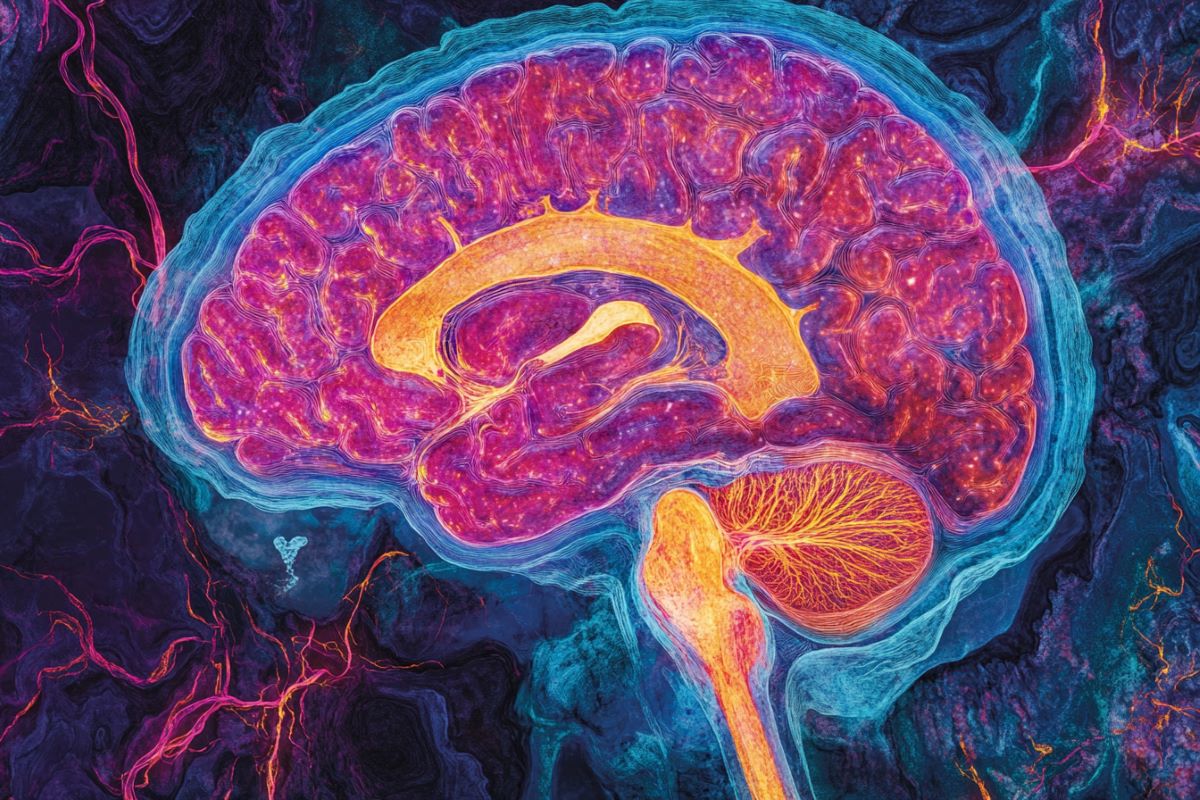
In the new study, the research team found that the motor cortex – the brain region responsible for controlling movement – becomes essentially “disconnected” during dyskinetic episodes.
This finding challenges the prevailing view that the motor cortex actively generates these uncontrollable movements.
Because of the disconnect between motor cortical activity and these uncontrollable movements, there’s probably not a direct link, but rather an indirect way in which these movements are being generated, Vishwanath said.
The researchers recorded activity from thousands of neurons in the motor cortex.
“There are about 80 billion neurons in the brain, and they hardly shut up at any point. So, there are a lot of interactions between these cells that are ongoing all the time,” Vishwanath said.
The research group found that these neurons’ firing patterns showed little correlation with the dyskinetic movements, suggesting a fundamental disconnection rather than direct causation.
“It’s like an orchestra where the conductor goes on vacation,” said Stephen Cowen, senior author of the study and an associate professor in the Department of Psychology.
“Without the motor cortex properly coordinating movement, downstream neural circuits are left to spontaneously generate these problematic movements on their own.”
This new understanding of dyskinesia’s underlying mechanism is complemented by the team’s findings regarding the therapeutic potential of ketamine, a common anesthetic.
The research demonstrated that ketamine could help disrupt abnormal repetitive electrical patterns in the brain that occur during dyskinesia. This could potentially help the motor cortex to regain some control over movement.
Ketamine works like a one-two punch, Cowen said. It initially disrupts these abnormal electrical patterns occurring during dyskinesia.
Then, hours or days later, ketamine triggers much slower processes that allow for changes in the connectivity and activity of brain cells over time, known as neuroplasticity, that last much longer than ketamine’s immediate effects.
Neuroplasticity is what that enables neurons to form new connections and strengthen existing ones.
With one dose of ketamine, beneficial effects can be seen even after a few months, Vishwanath said.
These findings gain additional significance in light of an ongoing Phase 2 clinical trial at the U of A, where a group of researchers from the Department of Neurology are testing low doses of ketamine infusions as a treatment for dyskinesia in Parkinson’s patients.
Early results from this trial appear promising, Vishwanath said, with some patients experiencing benefits that last for weeks after a single course of treatment.
Ketamine doses could be tweaked in a way such that the therapeutic benefits are maintained with minimized side effects, Cowen said. Entirely new therapeutic approaches may also be developed based on the study’s findings about motor cortex involvement in dyskinesia.
“By understanding the basic neurobiology underlying how ketamine helps these dyskinetic individuals, we might be able to better treat levodopa-induced dyskinesia in the future,” Cowen said.
Funding: The study received funding from National Institute of Neurological Disorders and Stroke (grants R56 NS109608 and R01 NS122805) and Arizona Biomedical Research Commission (grant ADHS18-198846).
About this Parkinson’s disease and neuropharmacology research news
Author: Niranjana Sahasranamam Rajalakshmi
Source: University of Arizona
Contact: Niranjana Sahasranamam Rajalakshmi – University of Arizona
Image: The image is credited to Neuroscience News
Original Research: Open access.
“Decoupling of motor cortex to movement in Parkinson’s dyskinesia rescued by sub-anaesthetic ketamine” by Abhilasha Vishwanath et al. Brain
Abstract
Decoupling of motor cortex to movement in Parkinson’s dyskinesia rescued by sub-anaesthetic ketamine
Gamma band and single-unit neural activity in primary motor cortex (M1) are involved in the control of movement.
This activity is disrupted in Parkinson’s disease (PD) and levodopa-induced dyskinesia (LID), a debilitating consequence of dopamine replacement therapy for PD. Physiological features of LID include pathological narrowband gamma oscillations, finely tuned gamma (FTG), and altered M1 firing activity.
Since most studies characterize LID through visual scoring, little is known about the relationships between ongoing dyskinetic movements, gamma, and neuronal activity at fast (sub-second) and slow (seconds) timescales.
Here, we investigate how motor cortex activity changes with movement at multiple timescales in animal models of PD and LID. Furthermore, sub-anesthetic ketamine has emerged as a possible therapy for LID.
How ketamine may reduce LID is not fully understood. Consequently, we investigate how ketamine affects the relationship between motor cortex activity and movement.


