Summary: A new study reports the larger the growth of areas associated with thinking in the cortex, the slower the growth of regions associated with emotional, sensory and motor skills.
Source: NIH/NIMH.
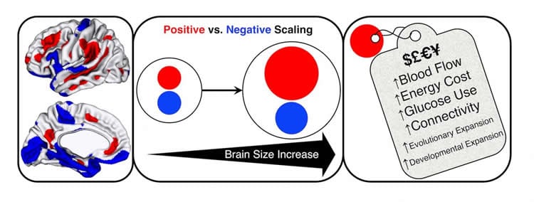
Some human brains are nearly twice the size of others – but how might that matter? Researchers at the National Institute of Mental Health (NIMH) and their NIH grant-funded colleagues have discovered that these differences in size are related to the brain’s shape and the way it is organized. The bigger the brain, the more its additional area is accounted for by growth in thinking areas of the cortex, or outer mantle – at the expense of relatively slower growth in lower order emotional, sensory, and motor areas.
This mirrors the pattern of brain changes seen in evolution and individual development – with higher-order areas showing greatest expansion. The researchers also found evidence linking the high-expanding regions to higher connectivity between neurons and higher energy consumption.
“Just as different parts are required to scale-up a garden shed to the size of a mansion, it seems that big primate brains have to be built to different proportions,” explained Armin Raznahan, M.D., Ph.D., of the NIMH Intramural Research Program (IRP). “An extra investment has to be made in the part that integrates information – but that’s not to say that it’s better to have a bigger brain. Our findings speak more to the different organizational needs of larger vs. smaller brains.”
Raznahan, Paul Reardon, Jakob Seidlitz, and colleagues at more than six collaborating research centers report on their study incorporating brain scan data from more than 3,000 people in Science. Reardon and Seidlitz are students in the NIH Oxford-Cambridge Scholars Program.
To pinpoint how the human brain’s organization varies in relation to how big it is, the researchers – including teams from the University of Pennsylvania, Philadelphia, and Yale University, New Haven, Connecticut, as well as the Douglas Mental Health University Institute, Verdun, Quebec – analyzed magnetic resonance imaging brain scans of youth from the Philadelphia Neurodevelopmental Cohort, a NIMH IRP sample, and the Human Connectome Project.
Cortex areas showing relatively more expansion in larger brains sit at the top of a network hierarchy and are specialized functionally, microstructurally and molecularly at integrating information from lower order systems. Since this theme holds up across evolution, development and inter-individual variation, it appears to be a deeply ingrained biological signature, Raznahan suggested.

“Not all cortex regions are created equal. The high-expanding regions seem to exact a higher biological cost,” explained Raznahan. “There’s biological ‘money’ being spent to grow that extra tissue. These regions seem to be greedier in consuming energy; they use relatively more oxygenated blood than low-expanding regions. Gene expression related to energy metabolism is also higher in these regions. It’s expensive, and nature is unlikely to spend unless it’s getting a return on its investment.”
Since people with certain mental disorders show alterations in brain size related to genetic influences, the new cortex maps may improve understanding of altered brain organization in disorders. The higher expanding regions are also implicated across diverse neurodevelopmental disorders, so the new insights may hold clues to understanding how genetic and environmental changes can impact higher mental functions.
“Our study shows there are consistent organizational changes between large brains and small brains,” said Raznahan. “Observing that the brain needs to consistently configure itself differently as a function of its size is important for understanding how the brain functions in health and disease states.”
“Notably, we saw the same patterns for scaled-up brains across three large independent datasets,” noted Seidlitz.
Funding: NIH/National Institute of Mental Health funded this study.
Source: Jules Asher – NIH/NIMH
Publisher: Organized by NeuroscienceNews.com.
Image Source: NeuroscienceNews.com image is credited to NIMH Developmental Neurogenomics Unit.
Original Research: Abstract for “Normative brain size variation and brain shape diversity in humans” by Katsuhiko Miyazaki, K. Reardon, Jakob Seidlitz, Simon Vandekar, Siyuan Liu, Raihaan Patel, M. T. M. Park, Aaron Alexander-Bloch, Liv S. Clasen, Jonathan D. Blumenthal, Francois M. Lalonde, Jay N. Giedd, Ruben Gur, Raquel Gur, Jason P. Lerch, M. Mallar Chakravarty, Theodore Satterthwaite, Russell T. Shinohara, and Armin Raznahan in Science. Published May 31 2018.
doi:10.1126/science.aar2578
[cbtabs][cbtab title=”MLA”]NIH/NIMH “Bigger Human Brain Prioritizes Thinking Hub, But at a Cost.” NeuroscienceNews. NeuroscienceNews, 1 June 2018.
<https://neurosciencenews.com/information-processing-priority-9210/>.[/cbtab][cbtab title=”APA”]NIH/NIMH (2018, June 1). Bigger Human Brain Prioritizes Thinking Hub, But at a Cost. NeuroscienceNews. Retrieved June 1, 2018 from https://neurosciencenews.com/information-processing-priority-9210/[/cbtab][cbtab title=”Chicago”]NIH/NIMH “Bigger Human Brain Prioritizes Thinking Hub, But at a Cost.” https://neurosciencenews.com/information-processing-priority-9210/ (accessed June 1, 2018).[/cbtab][/cbtabs]
Abstract
Normative brain size variation and brain shape diversity in humans
Brain size variation over primate evolution and human development is associated with shifts in the proportions of different brain regions. Individual brain size can vary more than twofold among typically developing humans, but the consequences of this for brain organization remain poorly understood. Using in vivo neuroimaging data from more than 3000 individuals, we find that larger human brains show greater areal expansion in distributed frontoparietal cortical networks and related subcortical regions than in limbic, sensory, and motor systems. This areal redistribution recapitulates cortical remodeling across evolution, manifests by early childhood in humans, and is linked to multiple markers of heightened metabolic cost and neuronal connectivity. Thus, human brain shape is systematically coupled to naturally occurring variations in brain size through a scaling map that integrates spatiotemporally diverse aspects of neurobiology.


