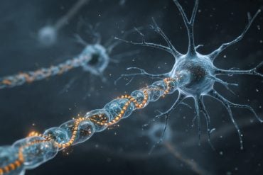
Summary: New research reveals that in people with ALS, structural changes in neurons activate immune cells, leading to inflammation and reduced motor function. A study explores the correlation between neuron changes and immune responses.
The findings suggest that blocking inflammation with a semi-synthetic drug derived from the Ashwagandha plant can restore synaptic connections, offering a potential treatment for ALS. This approach also holds promise for other inflammation-related diseases like Alzheimer’s.
Key Facts:
- ALS is characterized by the loss of upper and lower motor neurons, leading to muscle atrophy and motor function loss.
- Structural changes in upper neurons trigger immune responses, which become toxic if prolonged.
- A semi-synthetic drug based on Withaferin A can block inflammation and promote the regeneration of synaptic connections in motor neurons.
Source: Université Laval
In people with amyotrophic lateral sclerosis (ALS), changes in neurons appear to activate immune cells. Lowering the inflammation could reduce the symptoms of the disease, according to a study led by Chantelle Sephton, a professor at Université Laval’s Faculty of Medicine.
ALS is caused by the loss of upper motor neurons, located in the brain, and lower motor neurons, which extend from the spinal cord to the muscles. Using a genetically modified mouse model, Chantelle Sephton and her team found that structural changes in the upper neurons occurred prior to disease symptoms.
The study suggests that these morphological changes send a signal to microglia and astrocytes, the immune cells of the central nervous system. When they arrive, their effect is protective, but if they stay too long, they become toxic to neurons.
This leads to a reduction in synaptic connections between motor neurons in the brain and spinal cord, which in turn results in a reduction in synaptic connections with muscles. These changes lead to atrophy and loss of motor function.
Given this correlation between symptoms and immune response, the research team wondered whether it might be possible to restore synaptic connections by blocking inflammation.
” We tested a semi-synthetic drug based on Withaferin A, an extract of the Ashwagandha plant, which has been used for thousands of years in traditional Indian medicine,” explains CERVO Research Center affiliate Chantelle Sephton.
The drug blocks inflammation and allows motor neurons to return to a more normal state.
“We have noticed that neurons regenerate in the absence of activated immune cells. The dendrites of motor neurons start to grow and make connections again, increasing the number of synapses between motor neurons and muscles,” reports the researcher.
This seems a promising way of improving ALS symptoms, whether the disease is familial or sporadic, since both types are associated with inflammation.
Other diseases where inflammation plays a role, such as Alzheimer’s, could benefit from this approach.
About this ALS research news
Author: Audrey-Maude Vézina
Source: Université Laval
Contact: Audrey-Maude Vézina – Université Laval
Image: The image is credited to Neuroscience News
Original Research: Open access.
“Neuronal dysfunction caused by FUSR521G promotes ALS-associated phenotypes that are attenuated by NF-κB inhibition” by Chantelle Sephton et al. Acta Neuropathologica Communications
Abstract
Neuronal dysfunction caused by FUSR521G promotes ALS-associated phenotypes that are attenuated by NF-κB inhibition
Amyotrophic lateral sclerosis (ALS) and frontotemporal dementia (FTD) are related neurodegenerative diseases that belong to a common disease spectrum based on overlapping clinical, pathological and genetic evidence. Early pathological changes to the morphology and synapses of affected neuron populations in ALS/FTD suggest a common underlying mechanism of disease that requires further investigation.
Fused in sarcoma (FUS) is a DNA/RNA-binding protein with known genetic and pathological links to ALS/FTD. Expression of ALS-linked FUS mutants in mice causes cognitive and motor defects, which correlate with loss of motor neuron dendritic branching and synapses, in addition to other pathological features of ALS/FTD.
The role of ALS-linked FUS mutants in causing ALS/FTD-associated disease phenotypes is well established, but there are significant gaps in our understanding of the cell-autonomous role of FUS in promoting structural changes to motor neurons, and how these changes relate to disease progression.
Here we generated a neuron-specific FUS-transgenic mouse model expressing the ALS-linked human FUSR521G variant, hFUSR521G/Syn1, to investigate the cell-autonomous role of FUSR521G in causing loss of dendritic branching and synapses of motor neurons, and to understand how these changes relate to ALS-associated phenotypes.
Longitudinal analysis of mice revealed that cognitive impairments in juvenile hFUSR521G/Syn1 mice coincide with reduced dendritic branching of cortical motor neurons in the absence of motor impairments or changes in the neuromorphology of spinal motor neurons.
Motor impairments and dendritic attrition of spinal motor neurons developed later in aged hFUSR521G/Syn1 mice, along with FUS cytoplasmic mislocalisation, mitochondrial abnormalities and glial activation.


