

Summary: Study identifies the role the TMEM161B gene plays in excessive folding of the gyri in the cerebral cortex of those with polymicrogyria.
Source: UCSD
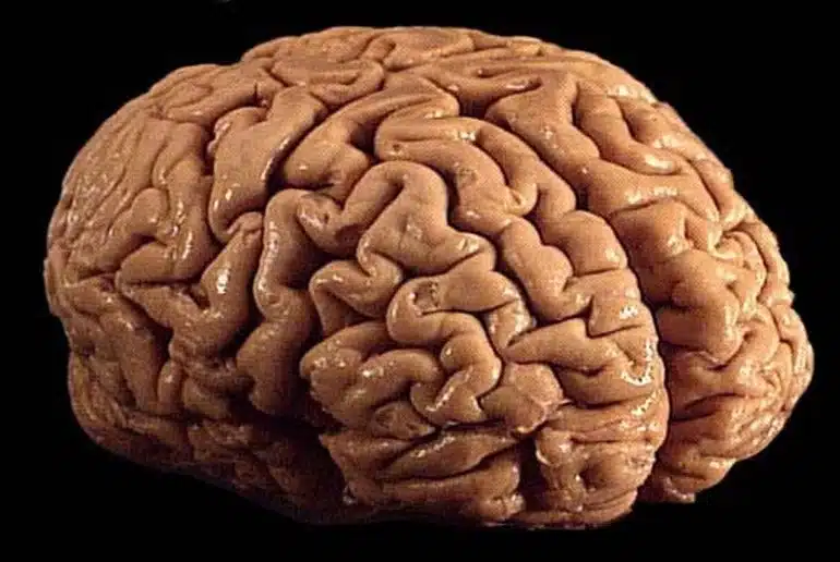
The outer layer of the human brain (the cerebral cortex), characterized by its distinctive gyri and sulci (those distinctive ridges and furrows), controls cognitive and executive function, from conscious thought to speech to emotional control.
The cerebral cortex is composed of more than 10 billion cells and 100 trillion-plus connections, a layer of gray matter just five millimeters thick—a little less than three stacked quarters.
Most animals with large brains exhibit cortical folding, which allows a very large area of cerebral cortex tissue (approximately 2.6 square feet) to be compacted inside the confines of the skull. The more cortical folding, the more advanced and complex the cognitive functions of the species.
Lower species like mice and rats have smaller, smooth surfaced brains; higher order species like elephants, porpoises and apes display different degrees of gyrification or folding of the cerebral cortex. Humans possess among the most wrinkly of brains, considered an indicator of advanced evolution.
In some humans, however, excess folding of the cerebral cortex is associated not with greater cognitive abilities, but the opposite and linked to neurodevelopmental delay, intellectual disability and epileptic seizures. The genes controlling this folding are mostly unknown.
Writing in the January 16, 2023 issue of PNAS, researchers at University of California San Diego School of Medicine and Rady Children’s institute for Genomic Medicine describe new findings that deepen understanding human gyrification.
Led by senior study author Joseph Gleeson, MD, Rady Professor of Neuroscience at UC San Diego School of Medicine and director of neuroscience research at the Rady Children’s Institute for Genomic Medicine, an international consortium of researchers called the Neurogenetics Consortium performed genomic analysis on nearly 10,000 families with pediatric brain disease over the course of 10 years to look for new causes of disease.
“From our cohort, we found four families with a condition called polymicrogyria, meaning too many gyri that are too tightly packed,” said Gleeson.
“Until recently, most hospitals treating patients with this condition did not test for genetic causes. The Consortium was able to analyze all four families together, which aided in our discovery of a cause for this condition.”
Specifically, all four families displayed mutations in a gene called Transmembrane Protein 161B (TMEM161B), which produces a protein of previously unknown function on cell surfaces.
“Once we identified TMEM161B as the cause, we set out to understand how excessive folding occurs,” said first author Lu Wang, Ph.D., a postdoctoral fellow in the Gleeson lab. “We discovered the protein controls the cellular skeleton and polarity, and these control folding.”
Using stem cells derived from patient skin samples, and engineered mice, the researchers identified defects in neural cell interactions early in embryogenesis.
“We found the gene is necessary and sufficient for cytoskeletal changes required for how neural cells interact with one another,” said Wang.
“It was interesting that the gene first appeared in evolution in sponges, which don’t even have a brain, so clearly the protein must have other functions. Here we found a critical role in regulating the number of folds in the human brain.”
The study authors emphasized that genetic discovery studies are important because they pinpoint causes of human disease, but that these discoveries can take many years to evolve into new treatments.
“We hope that physicians and scientists can expand upon our results to improve diagnosis and care of patients with brain disease,” said Gleeson.
About this neuroanatomy research news
Author: Scott LaFee
Source: UCSD
Contact: Scott LaFee – UCSD
Image: The image is credited to California Institute for Regenerative Medicine
Original Research: The findings will appear in PNAS


