Summary: A recent study reveals why most animals, including humans, inherit mitochondrial DNA solely from their mothers, showing that retaining paternal mitochondria can harm development and health. In roundworms, when paternal mitochondria persist, it disrupts ATP production, affecting neurological, behavioral, and reproductive health.
The study suggests that issues with paternal mitochondria clearance might play a role in certain mitochondrial disorders. Interestingly, treating affected worms with vitamin K2 restored ATP levels and improved their function, hinting at a potential new approach for managing mitochondrial diseases in humans.
Key Facts
- Paternal mitochondria can disrupt ATP production and harm health if retained.
- Vitamin K2 restored ATP levels in roundworms with compromised mitochondrial function.
- Study findings could inform future treatments for mitochondrial disorders.
Source: University of Colorado
It’s one of the basic tenets of biology: We get our DNA from our mom and our dad.
But one notable exception has perplexed scientists for decades: Most animals, including humans, inherit the DNA inside their mitochondria —the cell’s energy centers – from their mothers alone, with all traces of their father’s mitochondrial genome destroyed the moment sperm joins egg.
A new University of Colorado Boulder study published Oct. 4 in the journal Science Advances sheds new light on why this happens, showing that when the process fails, and paternal mitochondria slips into a developing embryo, it can lead to lasting neurological, behavioral and reproductive problems in adults.

The study, conducted in roundworms, offers new clues about what may drive some mitochondrial disorders, which hinder the body’s ability to produce energy and collectively impact about one in 5,000 people. It also presents a novel approach for potentially preventing or treating them – a simple vitamin known as Vitamin K2.
“These findings provide important new insights into why paternal mitochondria must be swiftly removed during early development,” said senior author Ding Xue, a professor in the Department of Molecular, Cellular and Developmental Biology (MCDB) at the University of Colorado Boulder.
“They also offer new hope for treatment of human diseases that may be caused when this process is compromised.”
When cellular batteries run low
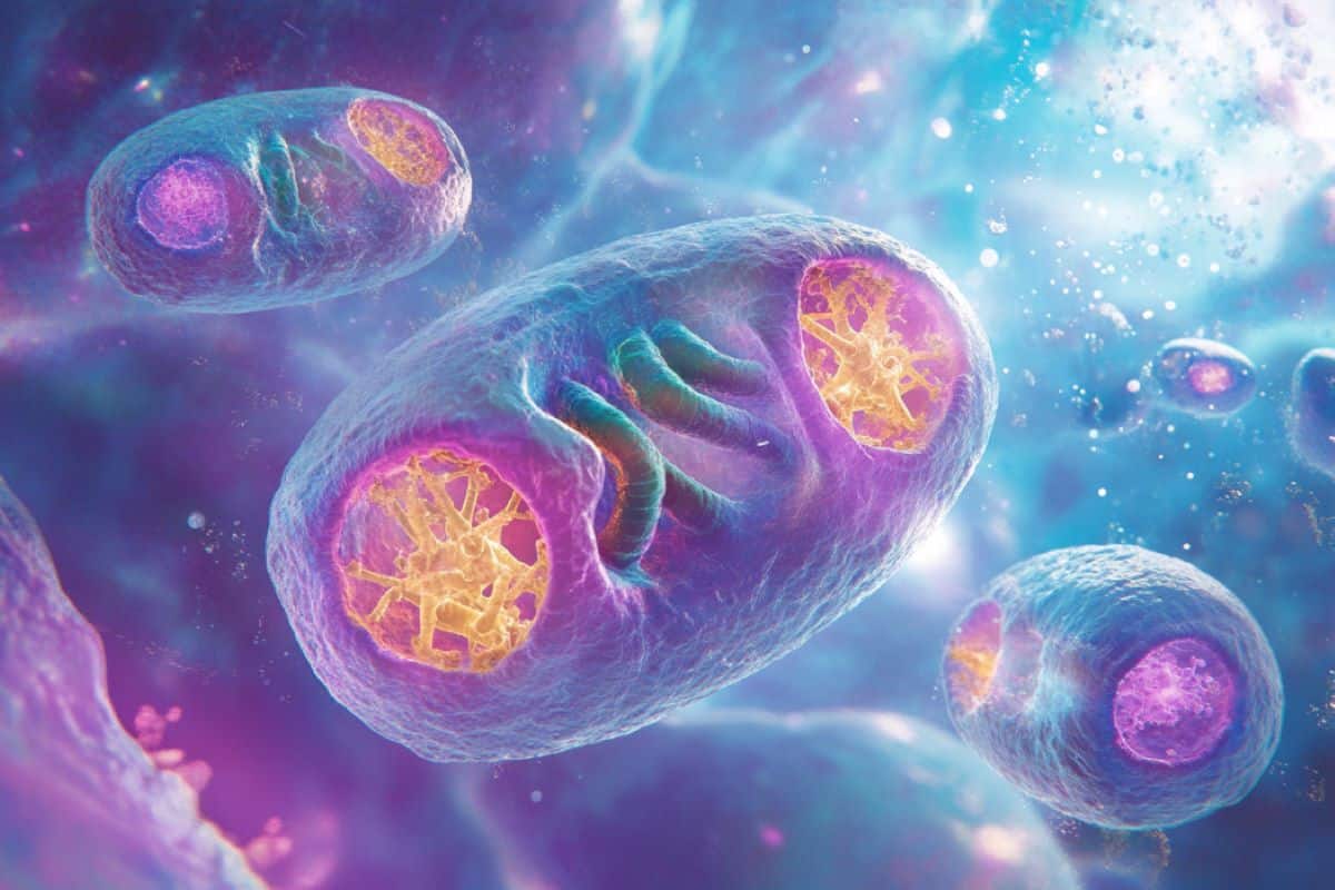
Often described as cellular batteries, mitochondria produce adenosine triphosphate (ATP), the energy that drives virtually all cell functions.
Mitochondria have their own distinct DNA, typically passed down exclusively from the mother.
In 2016, Xue published one of the first papers to spell out just how paternal mitochondria gets wiped out – via a multi-faceted, self-destruct mechanism known as “paternal mitochondria elimination (PME),” a process documented in worms, rodents and humans alike.
“It could be humiliating for a guy to hear, but it’s true,” Xue joked. “Our stuff is so undesirable that evolution has designed multiple mechanisms to make sure it is cleared during reproduction.”
Some have theorized that after battling it out with millions of other sperm to penetrate an egg, sperm mitochondria are exhausted and genetically damaged in ways that would be evolutionarily disastrous if passed on to future generations.
Xue and his team set out to find out what happens when paternal mitochondria do not self-destruct.
They studied C. elegans, a translucent worm which contains only 1,000 cells but develops a nervous system, gut, muscles and other tissues similar to humans.
The team was unable to completely halt PME in the worms – a testament to how resilient this evolutionary process is. But they were able to delay it by about 10 hours. When they did so in fertilized eggs, it led to significant reductions in ATP. If the worms survived at all, they had impaired cognition, altered activity and difficulty reproducing.
When the researchers treated the worms with a form of vitamin K2 known as MK-4 (best known as a bone health supplement) it restored ATP levels to normal in the embryos and improved memory, activity and reproduction in the adult worms.
Hope for little-understood diseases
The authors note that there are only a few documented cases in which paternal mitochondrial DNA might have been found in human adults. One paper describes a 28-year-old man had trouble breathing, weak muscles and could not tolerate exercise. Another documents 17 members of three unrelated multi-generational families who had fatigue, muscle pain, speech delays and neurological symptoms.
More research is needed in larger animals, but Xue suspects that in some cases, as with the worms, a mere delay in PME could be fueling hard-to-diagnose human diseases.
“If you have a problem with ATP it can impact every stage of the human life cycle,” he said.
Xue imagines a day when some families with a history of mitochondrial disorders take vitamin K2 prenatally as a precautionary measure. The study, and the lab’s ongoing research, could also lead to new ways to diagnose or treat mitochondrial disorders.
“There are a lot of diseases that are poorly understood. No one really knows what is going on. This research offers clues,” Xue said.
About this mitochondria and genetics research news
Author: Lisa Marshall
Source: University of Colorado
Contact: Lisa Marshall – University of Colorado
Image: The image is credited to Neuroscience News
Original Research: Open access.
“Moderate embryonic delay of paternal mitochondrial elimination impairs mating and cognition and alters behaviors of adult animals” by Ding Xue et al. Science Advances
Abstract
Moderate embryonic delay of paternal mitochondrial elimination impairs mating and cognition and alters behaviors of adult animals
Rapid elimination of paternal mitochondria following fertilization is a conserved event in most animals, but its physiological significance remains unclear.
We find that modest delay of paternal mitochondrial elimination (PME) in Caenorhabditis elegans embryos unexpectedly impairs mating and cognition of adult animals and alters their locomotion behaviors.
Delayed PME causes decreased adenosine triphosphate (ATP) levels in early embryos, which lead to impaired physiological functions of adult animals through an energy-sensing pathway mediated by an adenosine monophosphate (AMP)–activated protein kinase, AAK-2, and a forkhead box class O (FOXO) transcription factor, DAF-16.
Treatment of PME-delayed animals with MK-4, a subtype of vitamin K2 that can improve mitochondrial ATP production, restores ATP levels in early embryos, and rescues physiological defects of adult animals.
Our results suggest that moderate PME delay during embryo development adversely affects crucial physiological functions in adults, which could be evolutionarily disadvantageous.
These observations provide mechanistic explanations for the need to swiftly remove paternal mitochondria early during embryo development.


