

Summary: fMRI signals don’t always match the brain’s true activity levels, overturning a core assumption used in tens of thousands of studies. In about 40% of cases, an increased fMRI signal appeared in regions where neural activity was actually reduced, while decreased signals sometimes showed up in areas with heightened activity.
By measuring real oxygen use alongside fMRI, scientists found that many brain regions boost their efficiency by extracting more oxygen rather than increasing blood flow. These findings raise major questions about how brain disorders have been interpreted and suggest future imaging may need to shift toward direct measurements of energy consumption.
Key Facts:
- Mismatch Revealed: In roughly 40% of cases, higher fMRI signals were linked to lower neural activity.
- Oxygen Efficiency Shift: Brain regions often meet extra energy demand by extracting more oxygen instead of increasing blood flow.
- Clinical Impact: fMRI findings in depression, Alzheimer’s, and aging may reflect vascular differences rather than true neural activation changes.
Source: TUM
Researchers at the Technical University of Munich (TUM) and the Friedrich-Alexander-University Erlangen-Nuremberg (FAU) found that an increased fMRI signal is associated with reduced brain activity in around 40 percent of cases. At the same time, they observed decreased fMRI signals in regions with elevated activity.
First author Dr. Samira Epp emphasizes: “This contradicts the long-standing assumption that increased brain activity is always accompanied by an increased blood flow to meet higher oxygen demand. Since tens of thousands of fMRI studies worldwide are based on this assumption, our results could lead to opposite interpretations in many of them.”
Test tasks reveal deviations from the standard interpretation
PD Dr. Valentin Riedl, now Professor at FAU, and his colleague Epp examined more than 40 healthy participants during their time at TUM. Each was given several experimental tasks – such as mental arithmetic or autobiographical memory recall – which are known to produce predictable fMRI signal changes in distributed brain regions. During these experiments, the researchers simultaneously measured the actual oxygen consumption using a novel quantitative MRI technique.
Depending on the task and the brain region, the physiological results varied. Increased oxygen consumption – for instance in areas involved in calculation – did not coincide with the expected rise in blood flow.
Instead, the quantitative analyses showed that these regions met their additional energy demand by extracting more oxygen from the unchanged blood supply. Thus, they used the oxygen available in the blood more efficiently without requiring greater perfusion.
Implications for interpreting brain disorders
According to Riedl, these insights also affect the interpretation of research findings in brain disorders: “Many fMRI studies on psychiatric or neurological diseases – from depression to Alzheimer’s – interpret changes in blood flow as a reliable signal of neuronal under- or over-activation. Given the limited validity of such measurements, this must now be reassessed.
“Especially in patient groups with vascular changes – for instance due to aging or vascular disease – the measured values may primarily reflect vascular differences rather than neuronal deficits.” Previous animal studies already point in this direction.
The researchers therefore propose complementing the conventional MRI approach with quantitative measurements. In the long term, this combination could form the basis for energy-based brain models: rather than showing activation maps that depend on assumptions about blood flow, future analyses could display values indicating how much oxygen – and therefore energy – is actually consumed for information processing.
This opens new perspectives for examining aging, psychiatric, or neurodegenerative diseases in terms of absolute changes in energy metabolism – and for understanding them more accurately.
Funding: The research was conducted at the Neuro-Head Center of the Institute of Neuroradiology at the TUM University Hospital. It was funded by the European Research Council through an ERC Starting Grant.
Key Questions Answered:
A: Because fMRI relies on blood flow changes, not direct oxygen consumption, leading to misleading results when regions extract more oxygen from existing blood rather than increasing perfusion.
A: They combined fMRI with a quantitative MRI technique that directly tracked oxygen consumption, revealing discrepancies with standard blood-flow-based assumptions.
A: Many past studies may need reinterpretation, especially in groups with vascular aging or disease, where blood flow changes may not reflect neural function.
Editorial Notes:
- This article was edited by a Neuroscience News editor.
- Journal paper reviewed in full.
- Additional context added by our staff.
About this neuroimaging and neurotech research news
Author: Ulrich Meyer
Source: TUM
Contact: Ulrich Meyer – TUM
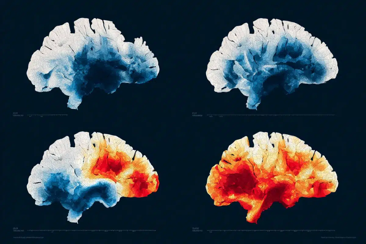
Image: The image is credited to Neuroscience News
Original Research: Open access.
“BOLD signal changes can oppose oxygen metabolism across the human cortex” by Valentin Riedl et al. Nature Neuroscience
Abstract
BOLD signal changes can oppose oxygen metabolism across the human cortex
Functional magnetic resonance imaging measures brain activity indirectly by monitoring changes in blood oxygenation levels, known as the blood-oxygenation-level-dependent (BOLD) signal, rather than directly measuring neuronal activity.
This approach crucially relies on neurovascular coupling, the mechanism that links neuronal activity to changes in cerebral blood flow. However, it remains unclear whether this relationship is consistent for both positive and negative BOLD responses across the human cortex.
Here we found that about 40% of voxels with significant BOLD signal changes during various tasks showed reversed oxygen metabolism, particularly in the default mode network.
These ‘discordant’ voxels differed in baseline oxygen extraction fraction and regulated oxygen demand via oxygen extraction fraction changes, whereas ‘concordant’ voxels depended mainly on cerebral blood flow changes.
Our findings challenge the canonical interpretation of the BOLD signal, indicating that quantitative functional magnetic resonance imaging provides a more reliable assessment of both absolute and relative changes in neuronal activity.


