

Summary: Researchers have identified a distinct excitatory neuron in the retrosplenial cortex that encodes direction related information over long durations.
Source: University of Michigan
It’s 5 p.m. as you leave the parking garage at work, but you realize you have no idea which way to turn to travel home. You know where you are and what street your house is on–it’s just that you can’t remember how to get there.
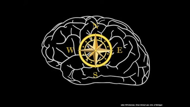
This is what happens to patients with damage to a part of their brain called the retrosplenial cortex, a key region involved in the organ’s inner compass. Despite its importance for navigation, the neurons and circuits it uses to help get people from the office to home remain understudied.
By recording signals from individual neurons in the mouse brain, researchers at the University of Michigan have identified a distinct excitatory neuron in the retrosplenial cortex. The properties of this neuron are ideally suited to encode direction-related information over long durations, like a compass.
“Regular neurons in the cortex are good at encoding directional information only when you are moving your head, but what happens when your head is still? You still need to know what direction you are facing so that you can use this information to plan your route,” said Omar Ahmed, assistant professor of psychology, neuroscience and biomedical engineering, and lead author of the study published in the journal Cell Reports.
“You ideally need another kind of neuron–a neuron that can continuously encode your orientation over long durations even when your head is not moving.”
Typical excitatory neurons slow down their firing rather quickly. In contrast, the newly identified neurons can continue firing their signals at high rates for extended periods of time–they are persistent and fast.
A second difference lies in their capacity to respond to inputs–these unique neurons, called low rheobase neurons–are hyperexcitable, which means they need little input to be activated.
“A simpler name for this small yet tenacious little neuron, as suggested by my classmate, would be ‘The Little Neuron That Could,'” said Ellen Brennan, the graduate student who identified these unique neurons. “It’s the perfect name because it highlights the persistence that makes them optimally suited to code continued direction. In comparison, the other typical excitatory neurons here are slow and stubborn.”
“So the question was, can these low rheobase neurons process directional information better than typical excitatory neurons?” said Shyam Sudhakar, a postdoctoral fellow in the Ahmed lab who created computer models of these neurons to show that the answer is “yes.”
“It’s important for my brain to know when I change direction, but it’s not good if all my brain detects is change,” Brennan said. “A compass always has to know which way is north. It wouldn’t be useful without that persistent sense of direction. That is exactly what the low rheobase neurons can provide.”
Credit: Omar Ahmed Lab.
Ahmed’s lab is now focused on understanding how these unique neurons are altered in Alzheimer’s model mice.
“The retrosplenial cortex is critical for spatial orientation, but is one of the earliest brain regions to show dysfunctional activity in Alzheimer’s patients,” Ahmed said. “This is probably why the vast majority of Alzheimer’s patients suffer from spatial disorientation and get lost easily–because their retrosplenial cells are not working as they should.
“By understanding how retrosplenial cells encode compass-like information in healthy versus Alzheimer’s brains, we hope to start working towards novel therapies.”
Other study co-authors include Izabela Jedrasiak-Cape and Tibin John, both members of Ahmed’s lab.
Source:
University of Michigan
Media Contacts:
Jared Wadley – University of Michigan
Image Source:
The image is credited to Omar Ahmed Lab.
Original Research: Open access
“Hyperexcitable Neurons Enable Precise and Persistent Information Encoding in the Superficial Retrosplenial Cortex”. Ellen K.W. Brennan, Shyam Kumar Sudhakar, Izabela Jedrasiak-Cape, Tibin T. John, Omar J. Ahmed.
Cell Reports doi:10.1016/j.celrep.2019.12.093.
Abstract
Hyperexcitable Neurons Enable Precise and Persistent Information Encoding in the Superficial Retrosplenial Cortex
Highlights
• Two distinct subtypes of excitatory neurons in superficial retrosplenial cortex (RSC)
• Most common neuron in layer 2/3 of RSC is excitatory low-rheobase (LR) neuron
• LR intrinsic properties enable precise, sustained encoding of information
• Layer 2/3 of RSC is dominated by feedforward, not feedback, inhibition
Summary
The retrosplenial cortex (RSC) is essential for memory and navigation, but the neural codes underlying these functions remain largely unknown. Here, we show that the most prominent cell type in layers 2/3 (L2/3) of the mouse granular RSC is a hyperexcitable, small pyramidal cell. These cells have a low rheobase (LR), high input resistance, lack of spike frequency adaptation, and spike widths intermediate to those of neighboring fast-spiking (FS) inhibitory neurons and regular-spiking (RS) excitatory neurons. LR cells are excitatory but rarely synapse onto neighboring neurons. Instead, L2/3 is a feedforward, not feedback, inhibition-dominated network with dense connectivity between FS cells and from FS to LR neurons. Biophysical models of LR but not RS cells precisely and continuously encode sustained input from afferent postsubicular head-direction cells. Thus, the distinct intrinsic properties of LR neurons can support both the precision and persistence necessary to encode information over multiple timescales in the RSC.


