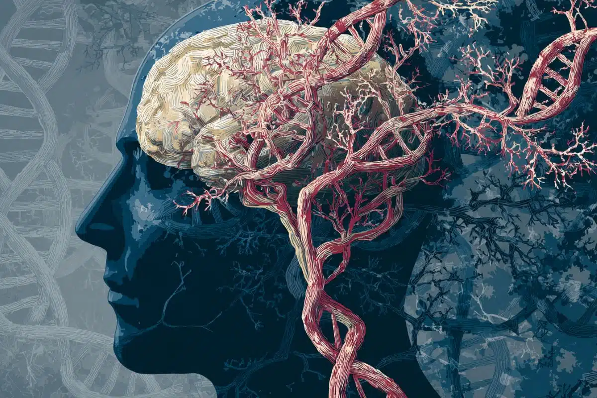
Summary: Chronic pain is often called the “invisible disability” because clinicians have no objective way to measure it—until now. A breakthrough study reveals that chronic pain has a unique “brain fingerprint” that differs radically from person to person.
By using intensive functional MRI (fMRI) longitudinal sampling on fibromyalgia patients over six months, researchers developed personalized AI models that can decode spontaneous pain fluctuations in real-time. The study proves that “one-size-fits-all” biomarkers won’t work for chronic pain; instead, the future of pain management lies in precision neuroimaging.
Key Facts
- The “Invisible” Becomes Visible: Using whole-brain functional connectivity, researchers successfully predicted moment-by-moment pain intensity without relying on a patient’s verbal report.
- Radical Individuality: The “pain connectome” identified in one patient was completely different from another. A model trained on Patient A could not predict the pain of Patient B, highlighting why chronic pain is so difficult to treat globally.
- Data Intensity Matters: The team found that standard, short-term brain scans are insufficient. Reliable “pain fingerprints” only emerged after extensive, repeated sampling over months.
- Fibromyalgia Focus: The study specifically looked at fibromyalgia, where pain often fluctuates spontaneously without an external trigger or injury.
- Precision Medicine: This research establishes a proof-of-concept for using non-invasive neuroimaging to evaluate pain objectively, which could eventually lead to personalized brain-targeted therapies.
Source: Institute for Basic Science
Chronic pain affects nearly one in five adults worldwide and remains one of the leading causes of disability. Unlike acute pain triggered by injury, chronic pain often arises spontaneously—without an obvious external cause—and fluctuates across minutes, hours, and days.
Yet clinicians still rely largely on self-reported pain ratings, as there is currently no objective biomarker comparable to blood pressure or body temperature.

Now, a research team led by Associate Director WOO Choong-Wan at the Center for Neuroscience Imaging Research (CNIR) within the Institute for Basic Science (IBS), in collaboration with Professor CHO Sungkun’s team at Chungnam National University, has demonstrated that personalized brain-imaging models can decode fluctuations in spontaneous pain intensity in individuals with chronic pain.
To address the challenge of capturing internally generated pain, the researchers conducted an intensive longitudinal study in patients with fibromyalgia, a chronic pain disorder characterized by widespread spontaneous pain.
Over more than half a year, participants underwent repeated functional magnetic resonance imaging (fMRI) sessions while continuously reporting their ongoing pain levels. fMRI measures changes in blood oxygenation across the whole brain, allowing researchers to examine patterns of communication between brain regions.
Using machine learning techniques applied to these densely sampled datasets, the team developed person-specific brain decoding models capable of predicting each participant’s moment-by-moment pain intensity.
The models successfully tracked fluctuations in spontaneous pain across multiple timescales—from minute-level changes within a scan to differences across sessions and days.
Importantly, prediction performance improved substantially as more training data were included. Conventional data quantities, typically used in longitudinal neuroimaging studies, were insufficient to achieve reliable predictions.
These findings highlight the importance of extensive within-person sampling when developing individualized brain-based biomarkers.
A key discovery of the study is that the neural patterns underlying pain differed markedly between individuals. The “pain connectome” identified in one participant did not generalize to another, and cross-testing between participants failed to produce meaningful predictions.
This individual specificity suggests that chronic pain is represented through highly personalized brain network configurations, underscoring the limitations of one-size-fits-all biomarkers.
Unlike earlier personalized decoding studies that relied on recordings from a limited number of brain regions, the present study leveraged whole-brain functional connectivity, capturing distributed network interactions known to be involved in pain processing.
The results demonstrate that spontaneous pain can be tracked using non-invasive neuroimaging, providing proof of principle for precision neuroimaging approaches in chronic pain research.
“The fact that pain cannot be seen adds to the suffering of patients with chronic pain,” said Dr. WOO Choong-Wan, Associate Director of IBS CNIR and senior author of the study. “Our findings show that precision neuroimaging may help evaluate invisible pain more objectively at the individual level.”
First author LEE Jae-Joong added, “Each participant exhibited a unique brain connectivity pattern associated with pain. Understanding these personalized neural signatures may eventually help guide precision approaches to pain assessment and treatment.”
Although the study involved a small number of participants and is not yet ready for clinical application, it establishes a methodological framework for developing patient-specific brain biomarkers.
Future research involving larger and more diverse cohorts will be necessary to determine whether subtypes of chronic pain share common neural features or require entirely individualized solutions.
Key Questions Answered:
A: This study shows that chronic pain isn’t just one signal in the brain—it’s a complex “conversation” between many different brain regions. Because every brain is wired differently, those conversations look unique to each person. What looks like “Level 8” pain in my brain might look like a totally different network pattern in yours.
A: Quite the opposite. This research provides objective, biological proof that chronic pain is a measurable physical state of the brain. It validates the suffering of millions of patients who have been told their pain isn’t “real” because it doesn’t show up on a traditional X-ray or blood test.
A: We aren’t there yet. This was a “proof-of-principle” study with a small group of patients. However, it provides the roadmap. The next step is scaling this technology so that it doesn’t require months of scans to build a personalized model.
Editorial Notes:
- This article was edited by a Neuroscience News editor.
- Journal paper reviewed in full.
- Additional context added by our staff.
About this AI and pain research news
Author: William Suh
Source: Institute for Basic Science
Contact: William Suh – Institute for Basic Science
Image: The image is credited to Neuroscience News
Original Research: Closed access.
“Personalized brain decoding of spontaneous pain in individuals with chronic pain” by Jae-Joong Lee, Seongwoo Jo, Sungkun Cho & Choong-Wan Woo. Nature Neuroscience
DOI:10.1038/s41593-026-02221-3
Abstract
Personalized brain decoding of spontaneous pain in individuals with chronic pain
Spontaneous pain is a hallmark of chronic pain disorders, but its assessment remains limited by the lack of objective biomarkers.
Here we used precision functional magnetic resonance imaging data, collected over more than half a year from two individuals with chronic pain, to develop personalized brain-decoding models of spontaneous pain.
The personalized decoding models accurately tracked fluctuations in spontaneous pain intensity across sessions, runs and minutes (Participant 1: prediction–outcome correlation, r = 0.40–0.61; Participant 2: r = 0.51–0.65) and effectively discriminated between median-dichotomized high- versus low-pain states (Participant 1: area under the curve = 0.71–0.87; Participant 2: area under the curve = 0.76–0.93).
Model performance improved with increased training data, with conventional data quantities failing to achieve significant predictive accuracy. Furthermore, each model relied on individually unique brain features and did not generalize across participants.
This study indicates that functional magnetic resonance imaging can assess spontaneous pain, highlighting the need for precise, patient-specific approaches.