

Summary: Study documents how CaMKII acts during synaptic plasticity, providing clues about the molecular signals that regulate memory formation.
Source: Max Planck Florida Institute For Neuroscience.
Researchers at Max Planck Florida Institute for Neuroscience optimized imaging methods to visualize the activation of CaMKII during synaptic plasticity with unprecedented resolution
Synaptic plasticity is the ability to strengthen or weaken the synapses or sites of communication between neurons. These changes are triggered by the activation of several different molecules inside small neuronal protrusions called dendritic spines. Although there are different forms of spine plasticity, these distinct forms are all induced by a common signal: the increase of calcium in the dendritic spines. The protein CaMKII is the most abundant protein kinase in the brain and is particularly enriched at synapses where it plays a crucial role in the regulation of synaptic transmission, neurotransmitter release, and synaptic plasticity. Normally, CaMKII is inactive because its binding site is blocked, but calcium removes the blockage, thus activating the protein. Communication through synapses causes calcium concentrations to spike, which activates CaMKII. But it is still a mystery exactly how calcium spike patterns translate into CaMKII activity.
In their May 2017 publication in Neuron, MPFI researchers from Ryohei Yasuda’s lab described how they optimized existing imaging methods to more precisely visualize CaMKII activation induced by calcium level increases. By improving the temporal resolution of fluorescence lifetime imaging to the millisecond range, they were able to see for the first time how CaMKII acts during synaptic plasticity in dendritic spines when calcium spikes occur in quick succession in a single dendritic spine. The researchers used laser pulses to activate single synapses and evoke calcium spikes in dendritic spines in mouse brain slices and then watched what happened to CaMKII activity. The Yasuda team found that CaMKII activity spiked in response to each pulse, just like calcium. But the CaMKII spikes lasted longer than the calcium spikes and combined together in a step-wise fashion. Because the signals combined over time, specific patterns of calcium spikes could translate into variable amounts of CaMKII activity and thus different changes in synapse strength and structure.
The protein CaMKII can also work independently of calcium if it phosphorylates itself at the site Thr286. This is because autophosphorylation prevents CaMKII’s binding site from becoming blocked again. Although this is an important feature of CamKII’s function, it is still unknown how phosphorylation influences CaMKII’s translation of calcium signals. To address this question, the Yasuda Lab repeated their experiments with a mutant form of CaMKII that couldn’t be phosphorylated at this site. The mutant CaMKII activity spikes were shorter than normal, and as a result, the activity induced by repeated synapse activation did not add up over time. Long-lasting changes in synapse strength and structure were also impaired. However, stimulating the synapse faster, so that the CaMKII spikes could add together, restored the functional and structural changes. These results indicate that Thr286 autophosphorylation is required for the induction of spine plasticity by facilitating calcium integration mediated by CaMKII, but phosphorylation is dispensable for the maintenance of the plasticity.
Future directions
This is the first report that demonstrates the precise filtering mechanisms of a calcium-dependent kinase during the induction of synaptic plasticity in single dendritic spines. The observations of this study clarify the critical role of Thr286 phosphorylation in plasticity: it lowers the stimulation frequency required to induce synaptic plasticity and permits CaMKII to integrate calcium signals at physiologically relevant frequencies. This provides direct evidence against the long-standing speculation that phosphorylation at Thr286 is the biochemical correlate for long-term memory.
Future research in the Yasuda Lab is expected to lead to significant advances in the understanding of the molecular signals that regulate memory formation. Because CaMKII function seems to be disturbed in mental diseases, a detailed analysis of this molecule using the advanced imaging methods developed in the Yasuda Lab may provide important insights into the pathogenesis of brain disorders, such as Alzheimer’s disease.
Funding: National Institutes of Health, Max Planck Florida Institute for Neuroscience, Max Planck Society funded this study.
Source: Jennifer Gutierrez – Max Planck Florida Institute For Neuroscience
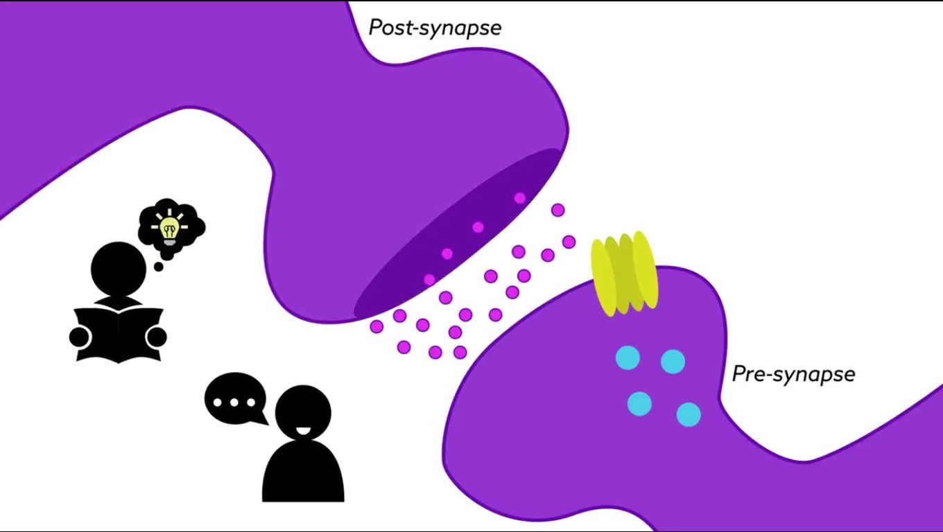
Image Source: NeuroscienceNews.com image is adapted from the Max Planck Florida Institute for Neuroscience video.
Video Source: Video credited to Max Planck Florida Institute for Neuroscience.
Original Research: Abstract for “CaMKII Autophosphorylation Is Necessary for Optimal Integration of Ca2+ Signals during LTP Induction, but Not Maintenance” by Jui-Yun Chang, Paula Parra-Bueno, Tal Laviv, Erzsebet M. Szatmari, Seok-Jin R. Lee, and Ryohei Yasuda in Neuron. Published online May 17 2017 doi:10.1016/j.neuron.2017.04.041
[cbtabs][cbtab title=”MLA”]Max Planck Florida Institute For Neuroscience “Precise Mechanisms of a Calcium Dependent Kinase During the Formation of New Memories.” NeuroscienceNews. NeuroscienceNews, 17 May 2017.
<https://neurosciencenews.com/calicum-kinase-memory-formation-6704/>.[/cbtab][cbtab title=”APA”]Max Planck Florida Institute For Neuroscience (2017, May 17). Precise Mechanisms of a Calcium Dependent Kinase During the Formation of New Memories. NeuroscienceNew. Retrieved May 17, 2017 from https://neurosciencenews.com/calicum-kinase-memory-formation-6704/[/cbtab][cbtab title=”Chicago”]Max Planck Florida Institute For Neuroscience “Precise Mechanisms of a Calcium Dependent Kinase During the Formation of New Memories.” https://neurosciencenews.com/calicum-kinase-memory-formation-6704/ (accessed May 17, 2017).[/cbtab][/cbtabs]
Abstract
CaMKII Autophosphorylation Is Necessary for Optimal Integration of Ca2+ Signals during LTP Induction, but Not Maintenance
Highlights
•CaMKII acts as a leaky integrator of Ca2+ pulses in dendritic spines
•Phosphorylation at T286 is necessary for optimal integration of Ca2+ pulses
•Phosphorylation at T286 is dispensable for the maintenance of LTP
Summary
CaMKII plays a critical role in decoding calcium (Ca2+) signals to initiate long-lasting synaptic plasticity. However, the properties of CaMKII that mediate Ca2+ signals in spines remain elusive. Here, we measured CaMKII activity in spines using fast-framing two-photon fluorescence lifetime imaging. Following each pulse during repetitive Ca2+ elevations, CaMKII activity increased in a stepwise manner. Thr286 phosphorylation slows the decay of CaMKII and thus lowers the frequency required to induce spine plasticity by several fold. In the absence of Thr286 phosphorylation, increasing the stimulation frequency results in high peak mutant CaMKIIT286A activity that is sufficient for inducing plasticity. Our findings demonstrate that Thr286 phosphorylation plays an important role in induction of LTP by integrating Ca2+ signals, and it greatly promotes, but is dispensable for, the activation of CaMKII and LTP.
“CaMKII Autophosphorylation Is Necessary for Optimal Integration of Ca2+ Signals during LTP Induction, but Not Maintenance” by Jui-Yun Chang, Paula Parra-Bueno, Tal Laviv, Erzsebet M. Szatmari, Seok-Jin R. Lee, and Ryohei Yasuda in Neuron. Published online May 17 2017 doi:10.1016/j.neuron.2017.04.041


