

Summary: MRI neuroimaging can distinguish between memory loss caused by Alzheimer’s disease and traumatic brain injury. Using new image analysis software, researchers discovered TBI causes the most amount of damage to the ventral diencephalon, a brain area associated with memory and learning, and the least amount of atrophy to the hippocampus, a brain region most impacted by Alzheimer’s.
Source: UCLA
Researchers from UCLA, along with colleagues at Washington University in St. Louis, say the finding is important because it could help prevent a misdiagnosis of Alzheimer’s disease, which can be devastating for patients and their families. One study found that as many as 21 percent of older adults with dementia may be misdiagnosed with Alzheimer’s disease. A misdiagnosis can result in patients not receiving the appropriate treatment, and prevents them from participating in clinical trials that could improve their overall care.
The current study, published in the Journal of Alzheimer’s Disease, involved 40 UCLA patients with an average age of just under 68, who had suffered traumatic brain injury, or TBI, and later developed memory problems. According to the U.S. Centers for Disease Control and Prevention 2.87 million Americans experienced TBI in 2014, with the rates highest for people age 75 or older. Children age 4 and younger, and adults age 65 and older were most likely to suffer serious brain injuries after a fall.
“We already knew that MRIs can reveal subtle abnormalities in patients with neurological disorders, such as Alzheimer’s,” said Dr. Somayeh Meysami, lead author and a postdoctoral clinical research fellow in cognitive and behavioral neurology at the David Geffen School of Medicine at UCLA. “The purpose of our study was to evaluate whether MRI also could reveal distinct abnormalities in traumatic brain injury. And, if we could identify such a pattern, it would lead to improved diagnosis of TBI-related memory loss from other causes of dementia.”
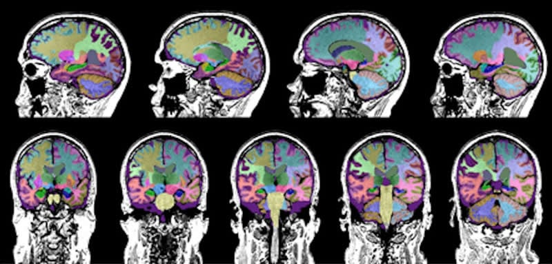
Using a software program to analyze the MRI scans, the study revealed that TBI caused the most damage to a brain region known as the ventral diencephalon, with the least amount of atrophy occurring in the hippocampus, said study author Dr. Cyrus Raji, an assistant professor of radiology at Washington University in St. Louis. The ventral diencephalon is associated with learning and emotions, whereas the hippocampus is involved in memory and emotions. The hippocampus also is the region of the brain that is most impacted by Alzheimer’s disease.
“The method we used to measure brain volumes in these individuals is useful because it can be applied on the same type of MRI scans we obtain in the clinic with no special type of imaging required,” Dr. Raji said.
The Alzheimer’s Association estimates that up to 40 percent of dementias are caused by conditions other than Alzheimer’s disease.
“Our study offers further evidence that not all memory loss is caused by Alzheimer’s disease,” he said. “It can attributed to TBI, as well as other dementias and neurodegenerative disorders,” said Dr. Mario Mendez, a professor-in-residence of neurology, psychiatry and biobehavioral Sciences at the David Geffen School of Medicine at UCLA.
Source:
UCLA
Media Contacts:
Marrecca Fiore – UCLA
Image Source:
The image is credited to UCLA Health.
Original Research: Open access
“Select this result for bulk action
MRI Volumetric Quantification in Persons with a History of Traumatic Brain Injury and Cognitive Impairment”. Somayeh Meysami et al.
Journal of Alzheimer’s Disease doi:10.3233/JAD-190708.
Abstract
Select this result for bulk action MRI Volumetric Quantification in Persons with a History of Traumatic Brain Injury and Cognitive Impairment
Background: While traumatic brain injury (TBI) is recognized as a risk factor for dementia, there is lack of clinical tools to identify brain changes that may confer such vulnerability. Brain MRI volumetric quantification can sensitively identify brain atrophy. Objective:To characterize regional brain volume loss in persons with TBI presenting with cognitive impairment.
Methods: IRB approved review of medical records in patients with cognitive decline focused on those who had documented TBI histories and brain MRI scans after TBI (n = 40, 67.7±14.5 years) with volumetric quantification by applying an FDA cleared software program. TBI documentation included head trauma mechanism. Brain volumes were compared to a normative database to determine the extent of atrophy. Correlations between these regions and global tests of cognition (MMSE in n = 17, MoCA in n = 27, n = 14 in both) were performed.
Results: Multiple regions demonstrated volume loss in TBI, particularly ventral diencephalon, putamen, and pallidum with smaller magnitude of atrophy in temporal lobes and brainstem. Lobar structures showed strongest correlations between atrophy and lower scores on MMSE and MoCA. The hippocampus, while correlated to tests of cognitive function, was the least atrophic region as a function of TBI history.
Conclusion: Persons with TBI history exhibit show regional brain atrophy. Several of these areas, such as thalamus and temporal lobes, also correlate with cognitive function. Alzheimer’s disease atrophy was less likely given relative sparing of the hippocampi. Volumetric quantification of brain MRI in TBI warrants further investigation to further determine its clinical potential in TBI and differentiating causes of cognitive impairment.


