Summary: A new study of the recently discovered brain’s lymphatic system may shed new light on various brain diseases and how fluid drainage may play a role.
Source: Rush University Medical Center
New research at RUSH is changing current understanding of the brain by revealing new knowledge about structures involved in cleansing it of fluids and waste with a steady fluid stream.
This system is called the brain’s lymphatic system—how fluid escapes the brain and drains into thin tubes—which allows for the circulation and removal of brain waste fluid.
The research is led by Rupal Mehta, MD, a physician-scientist and associate professor at RUSH University Department of Pathology and RUSH Alzheimer’s Disease Center.
Differing from current medical literature, the research shows that fluid escapes the human brain and drains into channels in the outer tissue layer that surrounds it. There are three thin layers of meningeal tissues in the outer leaflike covering that protects the brain.
This discovery is published online and in the February 2023 printed issue of the Journal of Experimental Medicine. New studies of this previously unknown area may help shed possible new clues about various brain diseases and what role it may play.
“In this study we describe arachnoid granulations, which are small mushroom-shaped outpouchings of the brain’s inner covering, as outlets that connect the brain plumbing system with adjacent lymphatic channels,” Mehta said.
“When leaving the brain, fluid and debris is carried away into arachnoid granulations. Therefore, granulations may teach us a whole lot regarding what’s going on in the brain, and how the brain functions and cleanses itself. Future studies on granulations may potentially help explain a range of brain diseases and lead to new therapeutics.”
The researchers found that a significant proportion of granulations border on bone and lymphatic channels or thin tubes, rather than the brain’s great veins as previous medical literature suggests. Medical literature describes arachnoid granulations as structures at the brain surfaces that drain passively and exclusively into the brain’s great veins called dural venous sinuses.
Before it was widely recognized that the brain had a lymphatic system, there was no reason to question arachnoid granulation structure. But with other recent discoveries in the field, Mehta and the RADC team decided to investigate for themselves.
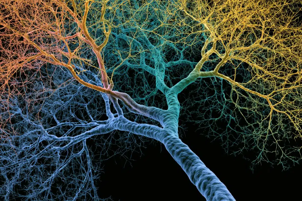
The authors analyzed more than 400 granulations across a number of brain specimens. Using a variety of microscopes and tissue preparation techniques, they examined the delicate tissues employing state-of-the art methods.
In recent years, scientists have described the glymphatic system, a fluid irrigation system within the brain that cleans out toxins. In combination with recently discovered lymphatic vessels present in the brain’s outer membranes, this system drains noxious materials from the brain.
This glymphatic system has been shown to carry brain waste from the brain, along the skull and to lymph nodes in the neck regions where the toxins are removed by the immune system.
Several studies now show that this brain drainage system plays important roles in a range of diseases. However, the fluid outlets, or region of glymphatic-lymphatic connection, in humans has been a mystery.
In the new study, the author Trishna Shah and colleagues used high-resolution microscopes with special staining and MRI techniques to characterize the shapes and arrangement of various parts of these granulation tissues in persons who varied in age.
The authors found that the internal framework of granulations consists of slivers of collagen that form a delicate sponge-like mesh or sieve, and contains different types of immune cells. The outer capsules of the granulations were also found to be perforated.
These findings differ from what is published in historical and modern-day textbooks. Moreover, the structure of granulations suggests that fluid and immune material percolate through the sponge-like tissues to escape onto the other side of the brain coverings, where the lymphatic vessels and bone are located.
“These structures appear to act like a filter that trap brain waste material and immune cells. They share several morphological features of peripheral lymph nodes,” Mehta said. “The findings suggest that the human brain has a well-developed and built-in mechanism for filtering and removing brain waste.”
The research shows how overlooked these tiny structures have been, but Mehta says that special high-power microscopes that were not widely available a few decades ago helped visualize the structure. Until now, detailed analyses using the newer microscopes have not been conducted. The functions of arachnoid granulations have not been fully understood by scientists and physicians.
For hundreds of years, they have been thought of as passive tissues and the brain coverings have been regarded as membrane-bound seals. Previously, it was not fully recognized that arachnoid granulations contain immune elements or communicate with spaces in the overlying tissues.
“This area of the brain is rarely analyzed and is overlooked on brain autopsy exams,” said Julie Schneider, co-author and director of the Alzheimer’s Disease Research Center. “Given that arachnoid granulations undergo significant aging changes, it could indicate a role in brain diseases like Alzheimer’s disease.”
In addition to unexpected location, shapes and arrangement of these parts, the authors also found a distinguishable age-associated change in arachnoid granulation content. The observations are important because they may fill in important gaps in what physicians and scientists know about brain aging and the unique ins and outs of the brain immune system.
Moreover, the findings could have importance for diseases associated with aging, such as stroke, trauma, and various forms of dementia. The authors hope that the discovery will translate into new insights for patients who suffer from neurological diseases. Mehta said that in future studies, the RADC team will study associations between arachnoid granulation deterioration and amyloid build-up in the brains of people with Alzheimer’s disease.
About this neurology research news
Author: Press Office
Source: Rush University Medical Center
Contact: Press Office – Rush University Medical Center
Image: The image is in the public domain
Original Research: Open access.
“Arachnoid granulations are lymphatic conduits that communicate with bone marrow and dura-arachnoid stroma” by Trishna Shah et al. Journal of Experimental Medicine
Abstract
Arachnoid granulations are lymphatic conduits that communicate with bone marrow and dura-arachnoid stroma
Arachnoid granulations (AG) are poorly investigated. Historical reports suggest that they regulate brain volume by passively transporting cerebrospinal fluid (CSF) into dural venous sinuses.
Here, we studied the microstructure of cerebral AG in humans with the aim of understanding their roles in physiology. We discovered marked variations in AG size, lobation, location, content, and degree of surface encapsulation.
High-resolution microscopy shows that AG consist of outer capsule and inner stromal core regions. The fine and porous framework suggests uncharacterized functions of AG in mechanical CSF filtration.
Moreover, internal cytokine and immune cell enrichment imply unexplored neuroimmune properties of these structures that localize to the brain–meningeal lymphatic interface. Dramatic age-associated changes in AG structure are additionally identified.
This study depicts for the first time microscopic networks of internal channels that communicate with perisinus spaces, suggesting that AG subserve important functions as transarachnoidal flow passageways.
These data raise new theories regarding glymphatic–lymphatic coupling and mechanisms of CSF antigen clearance, homeostasis, and diseases.